This is an automatically translated article.
The article was written by Anorectal gastroenterologist specialist, Robotic urological urological surgery unit Robot & Pediatric Surgery - Vinmec Times City International General Hospital.
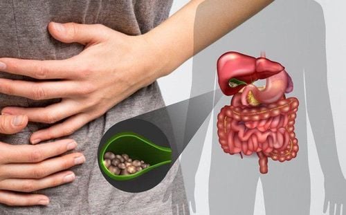
Acute cholecystitis due to gallstones is an inflammatory condition of the gallbladder, with obstruction caused by stones in the neck of the cystic duct or large stones that interfere with the normalization of the gallbladder with the biliary tree, acute cholecystitis is a leading surgical emergency in Vietnam. The disease is more common in women than in men, and occurs in middle-aged and elderly people.
1. Definition of acute cholecystitis
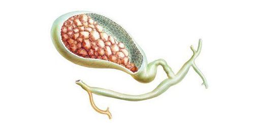
Causes of cholecystitis: 90% of acute cholecystitis have stones in the gallbladder. Stone stuck in the Hartman's funnel or cystic duct leads to stasis in the gallbladder -> inflammation, infection -> damage to the gallbladder mucosa.
In addition, it can be caused by mechanical friction of stones or chemical damage due to too high bile acid concentration, causing damage to the gallbladder mucosa.
Common pathogenic bacteria are anaerobic and anaerobic bacteria such as Streptococcus, Foecalis, Staphylococcus, E.Coli, Enterococci, Klebsiella, Bacteroides, Proteus...

2. Progression and forms of cholecystitis
Initiation: Stone stuck in the cystic duct causing congestive or exudative cholecystitis Inflammation of the gallbladder pustulitis: Following the process of congestive inflammation Necrotizing cholecystitis: When gallbladder inflammation leads to necrosis and perforation Gallbladder Peritoneal perforation: Bile fluid in the gallbladder seeps out of the abdomen Biliary peritonitis or perforation of the gallbladder Acute cholecystitis can form a clump of the gallbladder by the surrounding viscera, then inflammation Acute regression and subsequent formation of chronic cholecystitis Gallbladder cancer occurring on the background of gallstones or gallbladder polyps may present with chronic chronic cholecystitis.
3. Prognosis
Uncomplicated cholecystitis usually has a good prognosis, the mortality rate is quite low. Most cholecystitis, with appropriate treatment, resolves in 1-4 days. However, about 25-30% of cases do not respond to treatment, develop a complication of perforation necrosis requiring urgent surgical intervention.
Acute cholecystitis complicated by necrosis or perforation causing biliary peritonitis has a worse prognosis. Gallbladder perforation occurs in 10-15% of cases, with a mortality rate of about 5-10%.
4. Identification signs
4.1 Clinical Onset is biliary colic : Cramping pain in the right epigastrium or lower abdomen, may radiate to the right shoulder, pain usually occurs after a greasy meal ...
After more than 6 hours, the attack pain that does not subside may appear fever (38 or 38.5 degrees). If coming later, the patient has severe pain, reaction in the right lower quadrant on examination, higher fever (39-40 degrees), obvious infection syndrome: dry lips, dirty tongue, bad breath, fatigue, lethargy dull.
Some cases of cholecystitis may present additional jaundice. At that time, it is necessary to pay attention to the following problems: Cholecystitis accompanied by gallstones, cholecystitis with stones blocking the biliary tract (Mirrizzi's syndrome), cholecystitis with complications causing peritoneal biliary infiltration or peritonitis. bile , liver disease, etc.
In addition, vomiting may be accompanied by vomiting, or in rare cases there may be bowel obstruction or diarrhea, causing the physician to confuse the symptoms. other clinical conditions.
- Whole body
+ In the first stage, the infection syndrome appears with symptoms of high fever 38 - 39 degrees, chills intermittently; dry lips dirty tongue; bad breath.
+ The later stage is toxic syndrome: High fever, lethargy, panic (when the gallbladder has necrosis). Usually there is no obstructive jaundice. But when there are stones in the Hartmann's funnel (inserting into the right hepatic duct) or in the cystic duct (inserting into the common hepatic duct) causes obstructive jaundice (Mirizzi's syndrome).
In situ: The gallbladder is palpated in the right hypochondrium, a long, round, painful mass that moves with breathing, sometimes lying down to the pelvic cavity, lying on the skin of the abdomen (commonly seen). in the elderly and thin).

When examining the right subcostal area, there are signs of abdominal wall contracture or peritoneal tenderness.
4.2 Subclinical 4.2.1 Blood tests - Elevated white blood cells, increased neutrophil
- GOT, GPT, Total Bilirubin - direct, if there is an increase, attention should be paid to the problem of associated disease
- Amylase : If increased, it may be suspected of pancreatitis or not
- Other necessary tests to evaluate the patient: blood sugar, kidney function, coagulation function, blood group... and some other tests if clinical ready to have doubts.
Diagnostic imaging
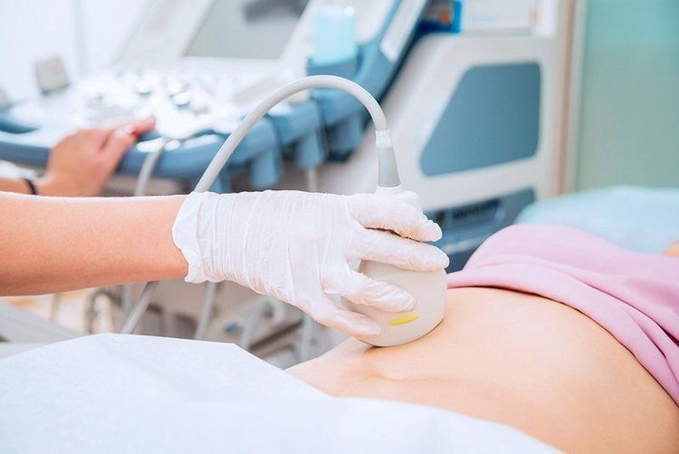
Ultrasound is an economical, but highly accurate, tool that is often used in the examination of acute abdominal pain. On the ultrasound image, the following information can be recorded: The gallbladder is distended and larger than normal (average length 8cm, transverse 3cm) inflammatory infiltrates, gallbladder wall thickness > 4mm, Image of the two sides of the wall of the pouch. gallstones or stones may be seen in the neck of the gallbladder. With or without fluid in the Morrison compartment.
X-ray of the abdomen upright and X-ray of the chest straight: To rule out the problems of perforation of the hollow viscera, intestinal obstruction, right lung inflammation.... sometimes the contrast image can be seen lying relative to the gallbladder position on the straight abdominal film or lean. CT scanning is indicated in difficult-to-diagnose or suspected cases of complicated cholecystitis or associated disease such as pancreatitis. MRI is also recommended for the diagnosis of acute cholecystitis, especially in patients with unclear symptoms or in pregnant women. Radioisotope imaging: Less recommended due to high cost. Gastroduodenal endoscopy may be indicated if the patient has responded to medical therapy.
Differential diagnosis
Usually requires differential diagnosis with the following conditions:
Coronary artery disease Acute pancreatitis Gastroduodenal ulcer Appendicitis Renal colic The following conditions should also be considered if weakness is present. Suspicion:
Abdominal aortic aneurysm Mesenteric ischemia Acute pyelonephritis Adnexitis

5. Treatment of acute cholecystitis
Acute cholecystitis is a delayed surgical emergency and the patient should be monitored in the Department of Surgery. Before implementing therapeutic measures, it is necessary to determine what stage of the acute inflammatory process the gallbladder is in. Inflammation of the gallbladder or necrosis of the gallbladder does not respond to medical therapy and emergency surgery is always indicated.
Once the diagnosis is confirmed, the treatment should be urgently implemented and include:
5.1 Active medical treatment Treatment principles include
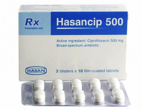
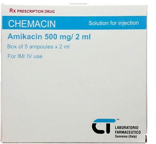
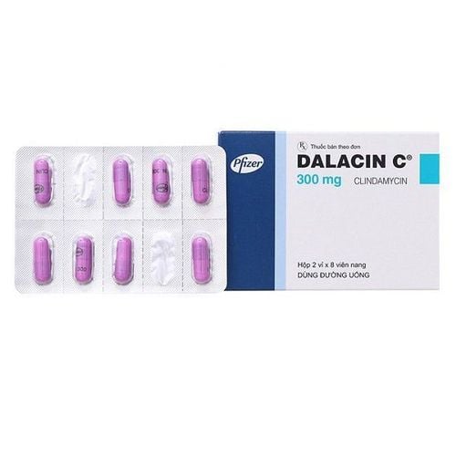
Fasting, possibly giving a nasogastric tube to Avoid irritating the gallbladder and pancreas. Nutritional infusions to rehydrate electrolytes, reduce secretions Use analgesics, anti-inflammatory drugs: NSAIDS, Perfalgan.... Inotropic or parasympathomimetic inhibitors: Atropine, nospa, alverine... Antibiotic treatment combination of third-generation cephalosporins with metronidazole. In severe cases of life-threatening inflammation, the antibiotic imipenem/cilastatin can be used

For patients with acute cholecystitis but no necrosis and perforation of the gallbladder, but have serious medical diseases that cannot be surgically intervened, can aspiration gallbladder drainage or open the gallbladder to the skin. .
If the patient is in good general condition with acute cholecystitis (ASA ≤ 2), emergency laparoscopic cholecystectomy will shorten the hospital stay.
5.2 Surgery Surgery should be started within the first 72 hours after antibiotics or earlier, open surgery or laparoscopic surgery are the two most commonly used methods of cholecystectomy due to acute cholecystitis.
Laparoscopic cholecystectomy is recommended in cases of stable endoscopically inflamed gallbladder and should be performed by experienced physicians.
The rate of laparoscopic conversion to open surgery in prepared cholecystectomy is about 5%. This rate is much higher in case of emergency surgery, about 30%.
Once diagnosed with complicated cholecystitis, it is necessary to operate immediately (laparoscopic or open surgery), or open the gallbladder to the skin if the patient is severe and cannot tolerate a long operation.
Perforation of the gallbladder causing localized abscesses (peri-gallbladder, subdiaphragmatic) or biliary peritonitis: pus culture is required for antibiotics, cholecystectomy with abdominal lavage and abdominal drainage.
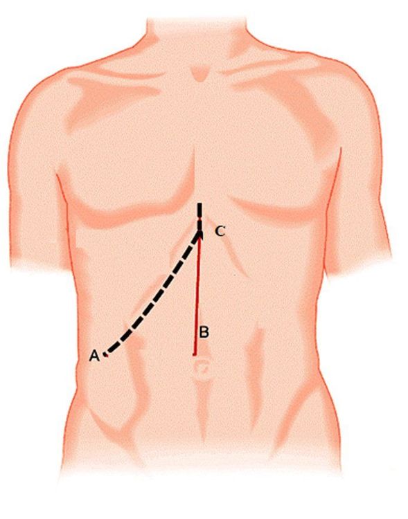
5.2.1. Open cholecystectomy The patients are under endotracheal anesthesia, the skin incision is usually applied: the incision follows the middle white line from the navel to the sternal nose or the horizontal line under the right ribs (rarely used).
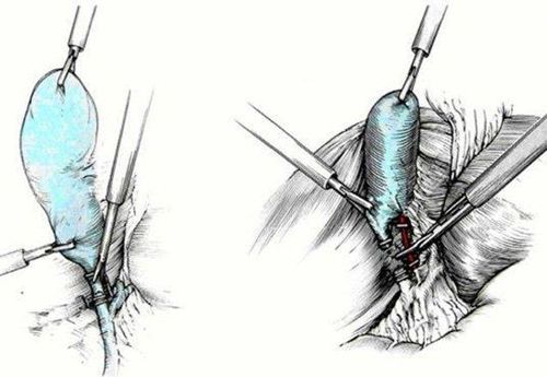
Dissection reveals the cystic duct and cystic artery. The cystic duct and cystic artery are managed with clips or sutures. Cholecystectomy from the gallbladder bed by unipolar electrocautery and histopathology. cystic artery and posterior cystic duct. After checking, if there is no damage to the liver, gastrointestinal tract, intestines, etc., a drainage can be placed under the right flank, ending the surgery.
5.2.2 . Laparoscopic cholecystectomy Similar to open surgery, the patient is under endotracheal anesthesia, lying supine. Manipulative channels are located in the umbilicus, below the ribs, and in the epigastrium. Using non-traumatic instruments, dissect the gallbladder and the area of the hepatobiliary triangle. Dissection revealed the cystic duct and cystic artery. The cystic duct and cystic artery are treated with clips or sutures, controlling the biliary tract to avoid injury to the biliary tract. Remove the gallbladder from the gallbladder bed with a unipolar electrocautery knife, carefully stopping the bleeding. For cases where it is difficult to identify the cystic duct, anterior cholecystectomy can be performed with a unipolar knife followed by ligation, cutting the cystic artery and posterior cystic duct. After the gallbladder is removed, a drainage can be placed under the liver to end the surgery.
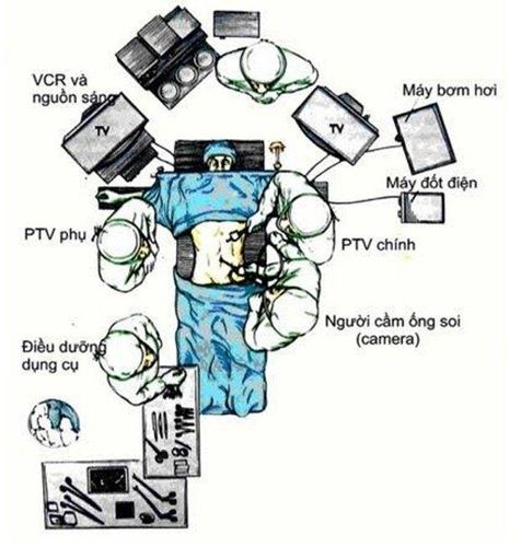
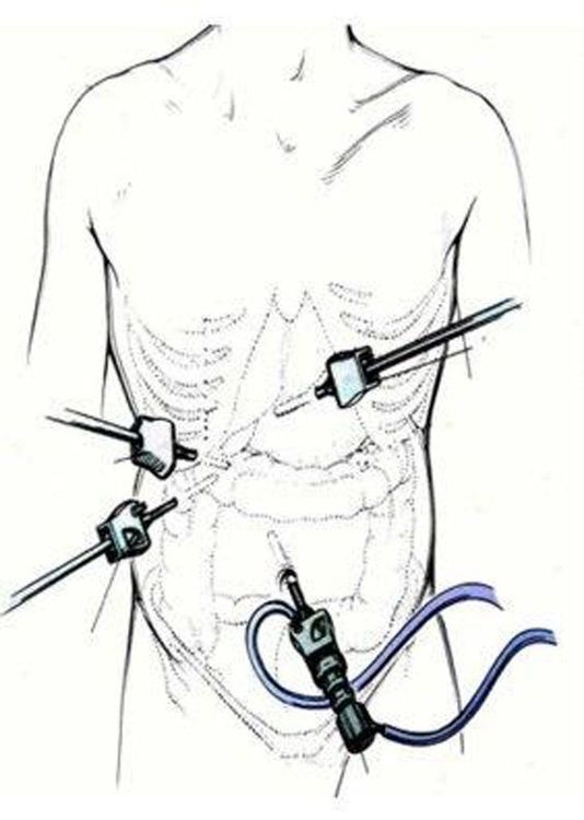
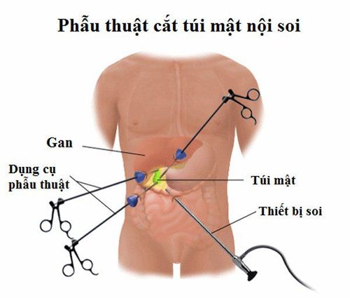
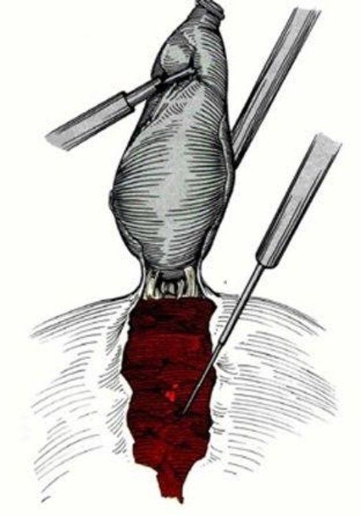
5.2.3. Complications and complications of surgery Biliary tract injury is the most feared complication. Therefore, laparoscopic cholecystectomy requires experienced surgeons and good dissection of Calot's triangle before resection, if in doubt, intraoperative cholangiography or switching to open surgery is required for safety.
Bleeding: Most bleeding comes from the gallbladder bed or from a small branch of the cystic artery. Bleeding is usually detected within 12 hours of surgery. This patient should be re-operated to resolve the bleeding site and obtain a blood clot.
Infection, residual abscess, fluid accumulation at the gallbladder bed, surgical site infection.
5.3 Post-operative treatment Post-operative antibiotics: Use parenteral or oral broad-spectrum antibiotics in combination with metronidazole. Postoperative analgesia: Infusion or oral administration of paracetamol in combination with tramadol, drugs in combination with drugs to reduce gastric secretion. Post-operative monitoring: Bleeding, pain, infection, biliary accident, complications of anesthesia: vomiting, dizziness, headache... Post-operative diet: Patients can eat porridge after surgery. 6 hours or at the discretion of the surgeon or anesthesiologist. After 6 hours of surgery, the patient can move lightly in bed and after 24 hours can walk and do normal activities. Drainage (if any) will be withdrawn after 3 days when there is no discharge or the amount of fluid is small and there is no abnormality Discharge: When the patient has no signs of bleeding, pain, infection, biliary stricture.. usually patients are discharged after 3-5 days of treatment Re-examination and suture removal after 07 days.

Note
All patients with acute cholecystitis should be hospitalized, monitored and treated at the Department of Surgery. All patients with gallstone cholecystitis should undergo cholecystectomy (elective or emergency surgery) unless surgery is contraindicated, requiring aggressive medical therapy. Cases of acute cholecystitis due to gallstones with indications for surgery need surgery soon for pain cases less than 10 days; In cases of pain over 10 days, surgery can be delayed after 30-45 days. Article referenced source:
A Treatment of acute calculous cholecystitis (uptodate Dec 2019 ) Laparoscopic cholecystectomy, CF142, V5, September 2013 Cambridge University Hospitals Cholecystitis ,emedicine.medscape 2011 Cho Ray Hospital's surgical treatment protocol (2013) . Acute cholecystitis. Cholecystitis. Pathology and treatment of gastrointestinal surgery (2007) Medical Publishing House of Ho Chi Minh City. Customers can directly go to Vinmec Health system nationwide to visit or contact the hotline here for support.


