This is an automatically translated article.
The article was professionally consulted by resident Doctor Nguyen Quynh Giang - Department of Diagnostic Imaging and Nuclear Medicine - Vinmec Times City International Hospital. Dr. Giang has many years of experience in the field of diagnostic imaging, especially in the field of multi-segment computed tomography, magnetic resonance.In most cases, when gallstones block the body's ducts out of the gallbladder, cholecystitis occurs. Other causes of cholecystitis include bile duct problems, tumors, serious illness, and some infections. To accurately diagnose the disease, doctors often use ultrasound, CT scan, and MRI techniques to evaluate gallbladder inflammation.
1. What is cholecystitis?
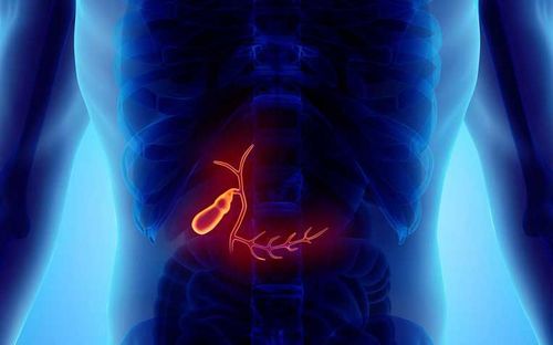
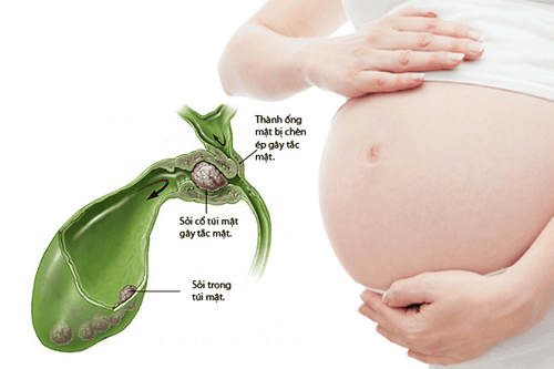
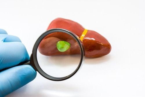
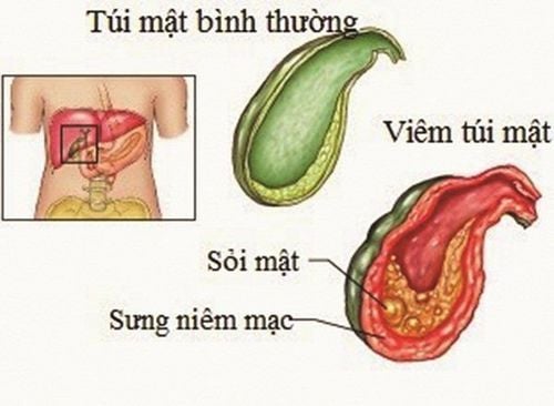
The gallbladder is a pear-shaped organ located under the body's liver and stores bile. If your gallbladder is inflamed, you may have pain in the upper right or middle part of your abdomen or it may be tender to the touch.Bile is made in the liver, the gallbladder stores bile and pushes it into the small intestine, where it is used to help digest food. When the drain for bile stored in the gallbladder (called the cystic duct) is blocked, usually by gallstones, the gallbladder becomes swollen and can become infected. This leads to cholecystitis. The cystic duct drains into the common bile duct, which carries bile into the small intestine. A gallstone can also become trapped in the common bile duct. This condition (gallstones) requires a procedure for removal.
Cholecystitis can be:
Acute cholecystitis (occurs suddenly): This inflammation often causes severe pain in the middle or upper right abdomen. Pain may also radiate between the shoulder blades. In severe cases, the gallbladder can tear or burst and release bile into the abdomen, causing severe pain. This can be a life-threatening situation that requires immediate attention. Chronic cholecystitis (multiple episodes of inflammation): Episodes of mild swelling and irritation, recurrent inflammation will often damage the gallbladder wall causing it to thicken, contract, and lose proper function. Cholecystitis can be caused by:
Gallstones : Cholecystitis is a common result of hard particles that develop in the body's gallbladder (gallstones). Gallstones can block the tube (cystic duct) through which bile flows as it leaves the gallbladder. Bile accumulates, causing inflammation.
2. Diagnostic and evaluation techniques for cholecystitis
Your doctor may order blood tests to see if you have a gallbladder infection. Normally, an elevated white blood cell count in our blood is a sign of an infection. However, for a more accurate diagnosis, your doctor may order one or more of the following optical tests:2.1. Ultrasound This is a routine test used to evaluate cholecystitis on ultrasound. Ultrasound uses sound waves to create images of the gallbladder and bile ducts. It is used to identify inflammatory markers related to the gallbladder and is very good at showing gallstones. Besides, ultrasound also has the advantages of easy implementation, low cost and high sensitivity.
Ultrasound image of acute cholecystitis is defined when the size of the gallbladder increases with the gallbladder transverse diameter greater than 4cm and the gallbladder wall thickness greater than 3mm. Fluid will be contained around the gallbladder and there are also indirect signs during ultrasound such as: positive ultrasound Murphy sign, increased flow on color Doppler ultrasound.
Furthermore, ultrasound of the gallbladder can also identify the causes and complications of acute cholecystitis such as: gallstones, gallstones, cholangitis or acute pancreatitis... Not only that, Gallbladder ultrasound also helps in differential diagnosis with other diseases such as liver diseases such as liver tumors, hepatitis, cirrhosis, ureteral stones, subhepatic appendicitis, ...
Some Patients should be advised to fast for 8 to 12 hours prior to performing an ultrasound evaluation of the gallbladder, because if eaten, the gallbladder will shrink and make examination and results difficult. Ultrasound results will not be accurate. It can also be the cause of missing small lesions.
2.2. Computed tomography (CT) This technique uses X-rays to create detailed images of the abdomen, liver, gallbladder, bile ducts, and intestines to help identify cholecystitis or obstruction of bile flow. Sometimes (but not always) it can also show gallstones.
2.3. Magnetic resonance cholangiography (MRCP) The MRCP technique is a type of MRI test that produces detailed images of the liver, gallbladder, bile ducts, pancreas, and pancreatic ducts. This technique is accurate in showing gallstones, cholecystitis or cholangitis, and blocked bile flow.
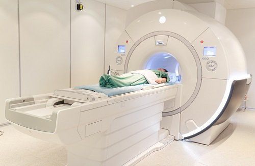
With many methods of diagnosing cholecystitis, it will help to treat the disease more effectively. However, these methods are expensive and time-consuming, so ultrasound-based testing is still considered the first clinical method used when screening for diseases related to ultrasound. gallbladder.
3. Treatment of cholecystitis
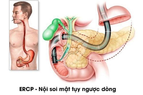
Treatment for cholecystitis usually includes a hospital stay to control the inflammation in your gallbladder. Sometimes, surgery is needed. In the hospital, your doctor will use a number of methods to control the signs and symptoms of the disease. Treatments may include:Fasting: You may not be allowed to eat or drink at first to relieve stress on the inflamed gallbladder Infusion of fluids through a vein: This helps prevent dehydration . Antibiotics to fight infection: If your gallbladder becomes infected, your doctor will likely recommend antibiotics. Pain relievers: These can help control pain until the inflammation in the gallbladder goes down. Procedure to remove stones: Your doctor may perform an endoscopic retrograde cholangiopancreatography (ERCP) procedure to remove any stones that block the bile duct or cystic duct. Symptoms of the disease are likely to subside in two or three days. However, cholecystitis often returns. Most people with the disease eventually need surgery to remove the gallbladder. Some procedures in the treatment of cholecystitis:
Percutaneous cholecystectomy: This procedure is done by a radiologist. The doctor will place a tube through the skin directly into the gallbladder using ultrasound. Blocked or infected bile is removed to reduce inflammation. This procedure is often done in patients who are too sick to have their gallbladder removed. You will be sedated for this procedure. The tubes usually should stay in the body for at least a few weeks. Endoscopic retrograde cholangiopancreatography (ERCP): This procedure is usually done by a doctor who specializes in abdominal disorders (gastroenterologist). A camera with a flexible tube is passed from the mouth through the stomach and into the beginning of the small intestine. This is where the common bile duct meets the small intestine. The valve mechanism (called a sphincter) at the end of the bile duct can be checked and opened to remove blocked bile and stones, if necessary. Doctors may also insert a small tube into the common bile duct and inject contrast material to better see the duct. They may also use fiber lasers to destroy small gallstones. All of this can be done without any incisions in the abdomen. You will be sedated during this procedure.
Please dial HOTLINE for more information or register for an appointment HERE. Download MyVinmec app to make appointments faster and to manage your bookings easily.
References: radiologyinfo.org, mayoclinic.org
SEE MORE
Treatment of cholecystitis by laparoscopic surgery What is the function of the gallbladder? Causes of cholecystitis Learn to cut cholecystitis due to gallstones by robotic laparoscopic surgery


