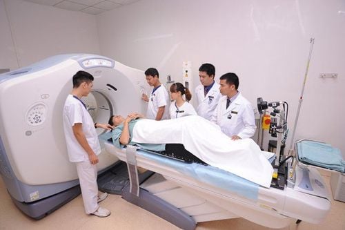
This is an automatically translated article.
This article is professionally consulted by Dr. Pham Quoc Thanh - Radiologist - Department of Diagnostic Imaging - Vinmec Hai Phong International General Hospital.1. Structure of veins
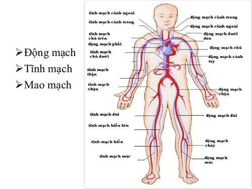
Veins are blood vessels in the circulatory system in the body that carry blood back to the heart (as opposed to arteries that carry blood from the heart) and when they have no capacity (for example, blood) they collapse. Using more blood, the veins will swell into tubes.The main structure of the vein consists of 3 main layers: the outer mantle, the middle mantle and the inner mantle. The outermost layer of veins is mainly composed of collagen surrounded by many rings of smooth muscle. The medial layer is thinner than the arterial medial layer, composed of circular smooth muscle fibers, less elastic fibers and collagen. The innermost layer of a vein is a layer of endothelial cells
The three layers of a vein are relatively thin and more dilated than an artery, so a vein can hold a relatively large amount of blood provided that the pressure is low. The internal force must change insignificantly. The main function of veins is to bring oxygen-poor and nutrient-poor blood back to the heart.
Veins play an extremely important role in the functioning of the body. Not only have the function of transporting, veins are also responsible for regulating our body and storing blood.
The venous system is located right next to our skin, so it can be easily observed with the naked eye, which is the superficial veins. As for the perforated veins, it will then assist in bringing blood to the deep veins. The muscles in our body will perform the main job is to pump to transport blood from the deep veins back to the heart.
2. When is an intravenous magnetic resonance imaging indicated?
When the patient is initially diagnosed with venous-related diseases such as venous thromboembolism,... When there is an abnormality of arteriovenous communication. Patients with congenital abnormalities related to veins, such as vascular malformations ... Follow the request from specialist doctors, assist in the diagnosis and treatment of a number of related diseases .3. Contraindicated to perform MRI without contrast injection in which cases?
Absolute contraindications:When the patient carries electronic devices such as: anti-vibration machine, machine specializing in cardiac rhythm regulation, device capable of automatically pumping drugs under the skin, cochlear implant, .. Patients using metal surgical forceps under the skin, intracranially, orbital, or vascular for < 6 months. Patients with complicated medical conditions must have resuscitation equipment by their side. Relative contraindications for the following cases:
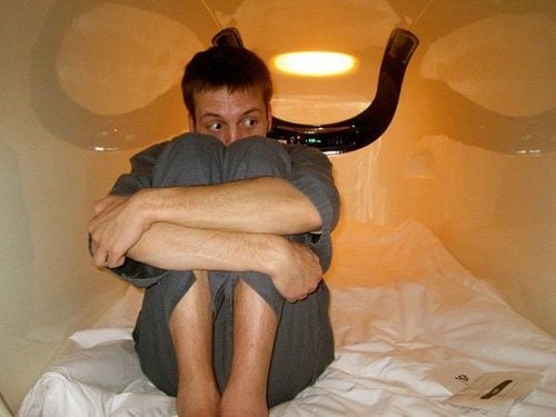
Use of metal surgical forceps for >6 months. Patients with claustrophobia, anxiety disorders,...
4. Necessary preparations before performing a non-magnetic contrast-enhanced intravenous MRI
4.1 Equipment The performer will be a specialist doctor and radiologist. The means used are magnetic resonance imaging machines of type 1.5Tesla or higher, film, film printers, together with a system with image storage function. Sedative . 4.2 Things to prepare for the patient The patient does not need to fast before the MRI scan. The doctor is responsible for consulting, answering questions, analyzing specifically the necessary procedures in the entire process so that the patient can coordinate well with the technician. Check for necessary contraindications. Instruct the patient to change the clothes of the MRI room and remove items not used during the scan. In order to perform an intravenous MRI, the patient should request a scan from the clinician with detailed diagnoses or specific medical records if needed.5. Magnetic resonance imaging (MRI) procedure from a vein without injecting contrast material
The patient lies on his back, installing equipment to receive peripheral pulse signals and receive signals from the whole body. In addition, the patient can wear a noise-cancelling headset if necessary.The imaging procedure is as follows:
Capture pulses of localized pulses of the area to be examined (lower extremities, upper extremities,...) in the form of three planes. Capture the pulse sequence of the vein according to the receiver of the peripheral pulse region. Reconstruct the plane (MPR for short) and the three-dimensional system (VRT). Evaluation of the results obtained:
Detect the lesions in the vein area if any. Images appear sharp with specific anatomical structures in the area examined. The specialist will detect abnormal lesions and describe them in a report on the internally connected computer and then print the results, thereby making accurate diagnoses. about the patient's current health status. Complications and treatment:
Patients with fear and agitation: psychological counseling, helping patients calm and reassure. Patients who are too nervous and scared before entering the MRI room due to claustrophobia may be advised to use sedation under the supervision of an anesthesiologist. Indications for intravenous magnetic resonance imaging without injection of magnetic contrast are necessary for early detection and diagnosis of dangerous diseases related to veins. This is a painless technique and affects the patient's health, so you can go directly to medical facilities equipped with an MRI room system for treatment advice and timely examination. .
In summary, this is one of the important techniques in imaging diagnosis to accurately assess the lesions and abnormalities of the venous system, which can replace Doppler ultrasound, computed tomography and digital imaging. background eraser. The scan time is about 45-60 minutes and the patient will receive the MRI results after 15-30 minutes, depending on the abnormalities in the body of the scanner.
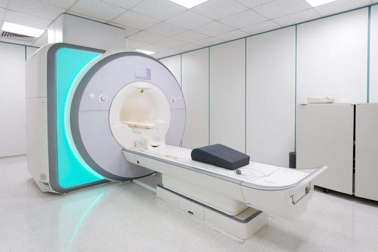
Vinmec International General Hospital put into use the magnetic resonance imaging machine 3.0 Tesla Silent technology. Magnetic resonance imaging machine 3.0 Tesla with Silent technology of GE Healthcare (USA).
Silent technology is especially beneficial for patients who are children, the elderly, weak health patients and patients undergoing surgery. Limiting noise, creating comfort and reducing stress for customers during the shooting process, helping to capture better quality images and shorten the shooting time. Magnetic resonance imaging technology is the technology applied in the most popular and safest imaging method today because of its accuracy, non-invasiveness and no use of X-rays. Doctor. Pham Quoc Thanh has received intensive training and participated in many national and international scientific conferences on diagnostic imaging. The doctor has 13 years of experience in the field of diagnostic imaging and is currently a doctor at the Department of Diagnostic Imaging, Vinmec Hai Phong International General Hospital.
Please dial HOTLINE for more information or register for an appointment HERE. Download MyVinmec app to make appointments faster and to manage your bookings easily.


