This is an automatically translated article.
The article is professionally consulted by Master, Doctor Pham Van Hung - Department of Medical Examination & Internal Medicine - Vinmec Danang International General Hospital. The doctor has 30 years of experience in examination and treatment of internal diseases, especially in Cardiology.1. How does the heart pump blood?
When it contracts, the heart pumps blood through a system of blood vessels called the circulatory system. Blood vessels are elastic muscular tubes that carry blood to every part of the body.Blood is an indispensable component for the life of the body. In addition to carrying fresh oxygen from the lungs and nutrients to the body's tissues, the blood also transports body wastes including carbon dioxide away from the body's tissues. This process is necessary to maintain life as well as promote the health of all parts of the body.
There are three main types of blood vessels :
Arteries . It begins with the aorta, which is the large artery that receives blood from the left chamber of the heart. Arteries carry oxygen-rich blood from the heart to all tissues of the body. They then branch out many times, getting smaller and smaller to transport blood from the heart to the organs of the body. Vascular . These are small, thin blood vessels that connect arteries and veins. The walls of the capillaries are thin to allow oxygen, nutrients, carbon dioxide and other waste products to easily enter or exit the cells of the body's organs. Vein . This is the blood vessel system that carries blood back to the heart. Venous blood has a lower oxygen content and contains more waste products to be eliminated or eliminated from the body. The size of the veins becomes larger as they get closer to the heart chambers. The superior vena cava is the large vein that carries blood from the head and arms to the heart. The inferior vena cava carries blood from the abdomen and legs back to the heart. The length of the vascular system (including arteries, veins, and capillaries) is more than 60,000 miles. With this length can be enough to circumnavigate the world more than twice.
2. Location and structure of the heart
The heart is located in the rib cage to the left of the breastbone and between the lungs. From the outside, it can be seen that the heart is made up of layers of muscle. These layers of heart muscle contract strongly to pump blood throughout the body.On the surface of the heart, there are coronary arteries, which supply oxygen-rich blood to the heart muscle itself. The main blood vessels that enter the heart are the superior vena cava, the inferior vena cava, and the pulmonary vein. The pulmonary artery leaves the heart and carries oxygen-poor blood to the lungs. The aorta carries oxygen-rich blood to the rest of the body.
Inside, the heart is an organ of 4 hollow chambers. The left and right heart chambers are divided by a muscular septum. The two upper chambers of the heart, called the atria, receive blood from the veins. The two lower chambers, called the ventricles, are where blood is pumped into the arterial system.
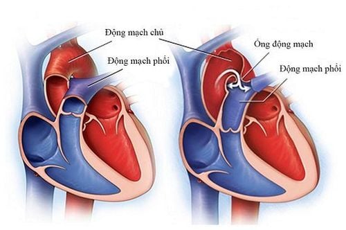
These valves work similarly to non-return valves in a water system. They prevent blood from flowing in the wrong direction. Each valve has a set of caps. Mitral valves have two flaps, while others have three flaps. These flaps are attached to and supported by a ring of sclerosing tissue called an annulus. These valve rings help maintain the proper shape of the valve.
The flaps of the mitral and tricuspid valves are also supported by tough, fibrous bands called fascia ligaments. This flap resembles a parachute, extending from the leaflets to the small muscles (papillary muscles) that are part of the inner wall of the ventricles.
3. The cycle of pumping blood through the heart
The right and left chambers of the heart work synergistically. This process repeats itself, causing blood to flow continuously to the heart, lungs, and body.3.1 Right Heart Blood enters the heart through two large veins, the inferior and superior vena cava, which carry oxygen-poor blood from the body into the heart's right atrium. When the atria contract, blood from the right atrium enters the right ventricle through the tricuspid valve.
When the ventricles are full, the tricuspid valve closes, which prevents blood from flowing back into the atrium during ventricular contraction. When the ventricles contract, blood leaves the heart through the pulmonary valve, into the pulmonary artery, and into the lungs. At the lungs, the blood is oxygenated and then returned to the left atrium through the pulmonary veins.
3.2. Left Heart The pulmonary veins carry oxygen-enriched blood into the left atrium of the heart. When the atria contract, blood flows from the left atrium into the left ventricle through the mitral valve.
When the ventricles are full, the mitral valve closes, which prevents blood from flowing back into the atrium during ventricular contraction. When the ventricles contract, blood leaves the heart through the aortic valve and enters the body.
4. The movement of blood through the lungs
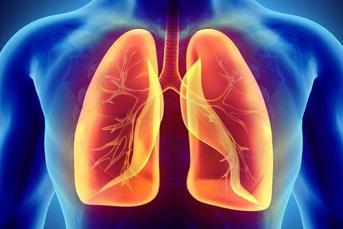
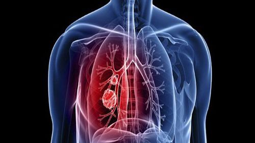
When blood passes through the pulmonary valve into the lungs, it is called pulmonary circulation. From the pulmonary valve, blood travels to the pulmonary artery and then to the small capillaries in the lungs. Here, oxygen from the alveoli in the lungs will pass through the capillary walls to enter the bloodstream. At the same time, carbon dioxide, which is a waste product of metabolism, passes from the blood into the alveoli. Carbon dioxide leaves the body through breathing. Once the blood has been purified and oxygenated, it returns to the left atrium through the pulmonary vein.5. Coronary artery of the heart
Like all organs, the heart is made up of tissues that require a supply of oxygen and nutrients. Although the chambers of the heart are filled with blood, the heart receives no nourishment from the blood. The heart receives its own blood supply from a network of arteries known as the coronary arteries.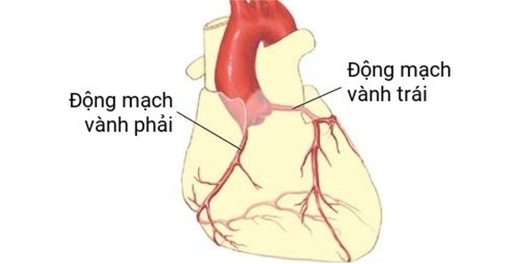
The left coronary artery consists of the circumflex artery and the anterior interventricular artery. The circumflex artery supplies blood to the left atrium, and posterior to the left ventricle; The anterior interventricular artery supplies blood to the anterior and inferior portions of the left ventricle as well as the anterior portion of the interventricular septum.
Coronary artery disease usually occurs when plaque builds up in the arteries that block the blood supply needed by the heart muscle, then a part of the small blood vessels in the heart will be blocked.
6. Heart activity
The atria and ventricles work together, alternately, and relax to pump blood through the heart. The heart's electrical system is the source of the energy that helps carry out this process.The heartbeat is triggered by electrical impulses traveling down from a special pathway through the heart. This electrical impulse begins in a small bundle of specialized cells called the sinus node located in the right atrium. This node is called a natural pacemaker. The electrical impulse propagates through the atrial septum and causes the atria to contract. A cluster of cells between the atria and ventricles is called the atrioventricular node - it's like a gate that slows down an electrical signal before it enters the ventricles. This delay gives the atria time to contract before the ventricles kick in.
The His-Purkinje network is a system of fibers that deliver electrical impulses to the interventricular septum and ventricular muscle, and cause the ventricles to contract.
At rest, the normal heart will beat about 50 to 99 times a minute. Heart rate can be faster than 100 beats/min with exercise, emotional changes, fever, and certain medications.
Vinmec International General Hospital is one of the hospitals that not only ensures professional quality with a team of leading medical professionals, modern equipment and technology, but also stands out for its examination and consultation services. comprehensive and professional medical consultation and treatment; civilized, polite, safe and sterile medical examination and treatment space. Customers when choosing to perform tests here can be completely assured of the accuracy of test results.
Please dial HOTLINE for more information or register for an appointment HERE. Download MyVinmec app to make appointments faster and to manage your bookings easily.
Article referenced source: Webmd.com