This is an automatically translated article.
The article is professionally consulted by MSc, BS. Dang Manh Cuong - Radiologist - Radiology Department - Vinmec Central Park International General Hospital. The doctor has over 18 years of experience in the field of ultrasound - diagnostic imaging.Computed tomography (CT-scanner) is a modern technique that uses 3-D images to capture the head and face, thereby helping doctors diagnose diseases deep inside the patient.
1. What is a 3D computed tomography (CT) scan of the brain?
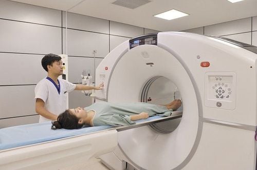
A CT scan of the brain, also known as a computed tomography scan of the brain, uses X-rays to take pictures of the head and face. This scan will provide pictures of the eyes, facial bones, the air spaces (sinuses) in the bones near the nose, and the inner ear. Therefore, cranial CT scan is used to evaluate diseases related to the head and face parts of the body.Computed tomography of the brain with 3D rendering is an advanced technique that helps surgeons locate in space the location of the lesion, thereby finding the fastest and safest approach to the lesion. 3D rendering techniques include 3D rendering of brain parenchyma, 3D of skull and 3D of cerebral blood vessels.
2. What diseases can a CT scan of the brain with 3D rendering detect?
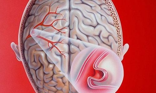
The doctor will conduct a CT scan of the brain when the patient has signs related to the brain, from which it is possible to detect the following diseases:Brain bleeding, find the cause of brain bleeding; Infarction in the presence of symptoms of a stroke (cerebral infarction); Locate brain tumors, soft tissue for treatment; Evaluation of ventricular dilatation , hydrocephalus ; Traumatic brain injury Diseases or congenital abnormalities of the skull and soft tissues; Stone pathologies in patients with hearing abnormalities; Assess for inflammation or other changes in the maxillary sinuses; Evaluation of cerebral vascular malformations such as aneurysms, arteriovenous catheters, ...; Guide needle biopsy for brain biopsies.
3. The outstanding advantages of cranial CT scan with 3D rendering
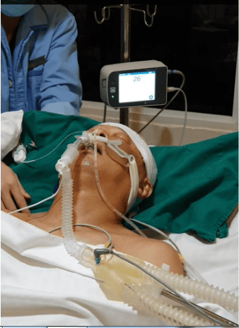
CT scan of the brain helps to accurately diagnose diseases related to the head and face. This is a modern and popular technique that is often prescribed by doctors if a patient is suspected of having a head injury with the following advantages:Evaluation of all tissues such as brain, bone, soft tissue, blood vessels in the same shot and for detailed images; Fast processing times should make sense in emergencies; Moderate cost per scan should be widely indicated in the clinical setting; Computed tomography is less affected by motion than magnetic resonance; Diagnostic computed tomography can help limit the rate of exploratory surgery and surgical biopsy.
4. Indication of cases requiring 3D computed tomography scan of the brain
Doctors will appoint the following cases to have 3D computed tomography scan:Suspected cases of cerebrovascular abnormalities such as subarachnoid hemorrhage, brain parenchymal bleeding, intraventricular bleeding .. Cerebral vascular malformations, cerebral aneurysms... Cases of skull subsidence, early craniosynostosis, skull deformity. Contrast foreign body in skull. Cases of brain tumors with indications for surgery or stereotactic radiation.
5. Contraindications during cranial computed tomography with 3D rendering
In the examination area, there are many metals that interfere with the image (relative contraindication). Pregnant patients (relative contraindications). There are contraindications to iodinated contrast agents.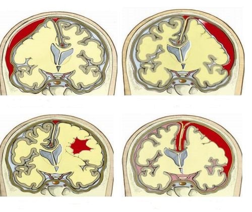
6. Steps in cranial computed tomography with 3D rendering
6.1. Place the patient
The patient is placed supine on the examination table. Move the table into the machine with the beam positioning for the examination area. Notes before taking the picture:Should wear loose shirt or depending on the place, there will be a separate shirt for the patient to wear before taking the picture; If your case requires contrast injection before the scan, you need to fast. At the same time, the doctor will take your history to see if you are allergic to any of the ingredients of the drug or have any special medical conditions and need to sign a consent form to inject contrast; Remove all metal items such as jewelry, eyeglasses, dentures, hairpins.
6.2. Shooting steps
Positioning imaging Set the cranial imaging field according to a process for the upper and lower examination areas of the tent Conduct for irradiation and image processing to evaluate the brain parenchyma obtained on the workstation screen, select the necessary images to reveal reveal pathology to print film. Place an vein with an 18G needle, connect a double-bore electric injection pump (1 barrel of medicine, 1 barrel of physiological saline). The usual amount of contrast agent used is 1.5 ml/kg body weight. Contrast is taken without contrast to clear the background. Perform a bolus test in the common carotid artery at the level of the C4 cervical vertebra. Select the time to take the X-ray during the injection, set the imaging field from C4 to the end of the skull. Carry out contrast injection and imaging, with physiological saline withdrawal. The acquired image will be modeled according to MIP, MPR, VRT programs to reveal the pathology. 3D modeling can be built in the shape of blood vessels, according to the brain parenchyma, according to the skull shape... The doctor reads the description on the internal connection computer and prints the results. 3D rendering of cerebrovascular system, brain parenchyma, skull clearly and fully. The skull is an important organ for every person. Therefore, you should pay attention to the signs of not being good about the brain for early detection and treatment. With a team of masters, doctors, technicians with deep expertise, high sense of responsibility and equipped with modern and synchronous machinery system, VINMEC hospital is committed to providing patients with excellent services. The best and most effective healthcare.Especially in order to improve diagnostic results, Vinmec has been and continues to invest in modern CT, X-ray, and MRI scanners... in combination with medical examination and treatment services. early treatment and control of cancers.
Please dial HOTLINE for more information or register for an appointment HERE. Download MyVinmec app to make appointments faster and to manage your bookings easily.


