This is an automatically translated article.
The article is professionally consulted by Master, Doctor Mai Vien Phuong - Gastroenterologist - Department of Medical Examination & Internal Medicine - Vinmec Central Park International General Hospital.The esophagus is the first part of the digestive tube that carries food from the pharynx to the stomach, about 25cm long, relatively mobile, attached to the surrounding organs by loose, flattened structures because the walls are close to it. each other and have a tubular shape when swallowing food.
Above the esophagus is connected to the pharynx at the level of the 6th cervical vertebrae, below the gastric outlet at the cardia, at the level of the 10th thoracic vertebrae. The esophagus is located posterior to the trachea, descends to the posterior mediastinum, is located posteriorly. posterior to the heart and anterior to the thoracic aorta, through the diaphragm into the abdomen, to the stomach.
1. Structure of the esophagus
Anatomically, the esophagus is about 25cm long and at its narrowest point is about 1.5cm in diameter divided into 3 segments: cervical; thorax and abdomen.
The lumen of the esophagus also has three narrow places: the junction with the pharynx at the level of the cricoid cartilage; at the level of the left aorta and left main bronchus; heart hole.
Neck segment: about 3cm long, food will run through the trachea and then to 1/3 of the neck, the food will shift to the left and down to run parallel to the trachea and then continue through the inlet behind the chest to enter the chest segment. Chest segment: about 20cm long, food entering the chest will continue through the diaphragm to enter the abdomen. Abdominal segment: about 2cm long, food has reached the abdomen, through the foramen into the stomach. Structurally, the esophagus is composed of skeletal and smooth muscle layers, the inner surface is covered by mucosa, the outermost layer is the connective tissue layer that surrounds the esophagus.
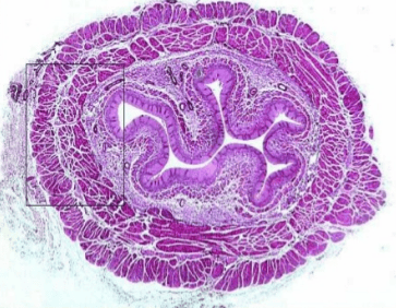
Các lớp của thực quản quan sát dưới kính hiển vi, lớp trong cùng là lớp niêm mạc thực quản, nơi dễ xảy ra bệnh lý viêm thực quản
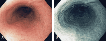
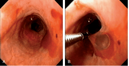
Hình ảnh nội soi của thực quản bình thường
2. What is esophagitis?
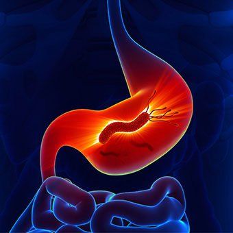
Esophagitis is a condition in which the lining of the esophagus is damaged and leads to inflammation. The most common causes of esophagitis are acid reflux, a side effect of certain medications, and bacterial or viral infections. Acid reflux occurs when the contents of the stomach back up into the esophagus.
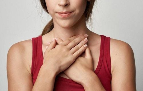
Common signs of esophagitis include:
Difficulty swallowing Sore throat Heartburn If left untreated, esophagitis can lead to esophageal ulcers, scarring, severe narrowing of the esophagus and need first aid.
But with prompt treatment, most cases improve significantly after about 2 to 4 weeks. However, in people with weakened immune systems or infections, treatment may take longer and recovery may take longer.
3. Types of esophagitis
3.1 Eosinophilic esophagitis
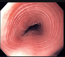
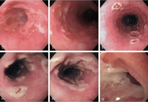
Viêm thực quản tăng bạch cầu ái toan với hình ảnh đặc trưng là các vòng tròn đồng tâm ở thực quản trên nội soi
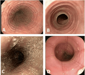
Một hình ảnh khác của viêm thực quản tăng bạch cầu ái toan
Eosinophilic esophagitis is caused by too many eosinophils in the esophagus. This happens when the body overreacts to an allergen. In children, this type of esophagitis makes eating difficult. Common disease triggers include:
Soy Milk Eggs Wheat Flour Peanuts Nuts Shellfish Inhalation allergens, such as pollen, are also among the common triggers. cause of this form of esophagitis.
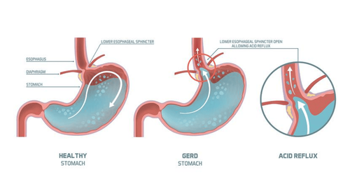
3.2 Reflux esophagitis Reflux esophagitis is usually caused by gastroesophageal reflux disease (GERD). Gastroesophageal reflux disease occurs when stomach contents, including acid, regularly back up into the esophagus, causing irritation and chronic inflammation of the lining of the esophagus.
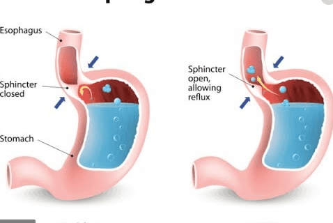
Viêm thực quản trào ngược xảy ra khi các chất trong dạ dày, bao gồm cả axit thường xuyên trào ngược lên thực quản
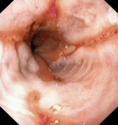
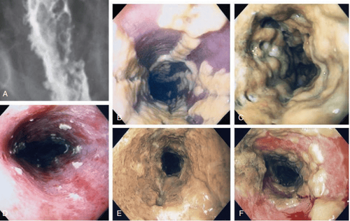
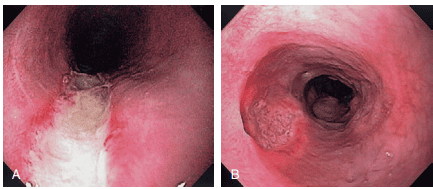
Một trường hợp viêm thực quản trào ngược được xác định trên nội soi
Viêm thực quản trào ngược với đặc trưng là các vết xước chạy lên từ chỗ nối dạ dày với thực quản
3.3 Drug-induced esophagitis Drug-induced esophagitis occurs when certain medications are taken without adequate fluids. Some oral medications can cause tissue damage if they remain in contact with the lining of the esophagus for a prolonged period of time.
For example, if a tablet is swallowed with little or no water, the tablets or residue from the tablet may remain in the esophagus. Medications that have been linked to esophagitis include:
Aspirin and other nonsteroidal anti-inflammatory drugs (NSAIDs) such as ibuprofen and naproxen. Antibiotics, such as tetracycline and doxycycline. Potassium chloride, used to treat potassium deficiency. Bisphosphonates, including alendronate, treat weak and fragile bones (osteoporosis).
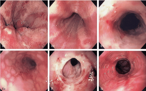
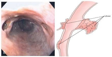
Một trường hợp bệnh nhân bị viêm thực quản do thuốc với các tổn thương mất niêm mạc và có giả mạc bám trên bề mặt
3.4 Infectious esophagitis Infectious esophagitis is very rare and can be caused by bacteria, viruses, fungi, or parasites. People at high risk for this type of esophagitis are people with weakened immune systems due to illness or medication. Infectious esophagitis occurs most commonly in people with HIV/AIDS, cancer, and diabetes.
Hình ảnh viêm thực quản do nấm
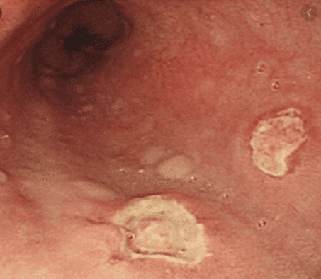
Hình ảnh viêm thực quản do virus Herpes Simplex
Viêm thực quản do Cytomegalovirus
Viêm thực quản do Cytomegalovirus
Viêm thực quản dạng bóng nước (bệnh Pemphigus)
Please dial HOTLINE for more information or register for an appointment HERE. Download MyVinmec app to make appointments faster and to manage your bookings easily.
Images in this article are taken at the source:
Cancertherapyadvisor.com Clinical Gastrointestinal Endoscopy by Hoon Jai Chun, Suk-Kyun Yang, Myung-Gyu Choi.