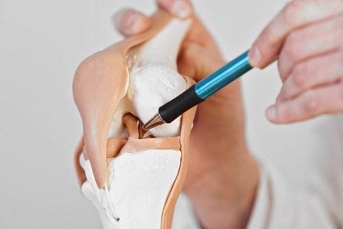
The meniscus is a rubber-like elastic cushion located between the femur and the tibia, acting as a shock absorber for the knee. There are two menisci, one medial, and one lateral. Of which, Grade 2 medial meniscus tears are often severe. The characteristic of Grade 2 medial meniscus tears is that they can be split in half or torn around the circumference in a C-shape and often require surgical intervention.
1. Anatomical characteristics of the medial meniscus
Similar to the lateral meniscus, the medial meniscus is also curved in shape with a length of about 3.5 cm. The anterior horn of the medial meniscus is attached to the front of the tibia and is far from the tibial plateau. The anterior fibers of the anterior cruciate ligament fuse with the transverse ligament connecting the anterior horns of the medial meniscus. The posterior horn of the medial meniscus is firmly attached to the back of the joint capsule. At the midpoint, the medial meniscus is attached to the femur and tibia by a ligament called the deep medial collateral ligament.
The vascular supply of the meniscus originates mainly from the medial, inferior, and superior geniculate arteries. During the first year of life, the meniscus is vascularized throughout its entire structure, but as the body ages, the vascular network diminishes and only 25-33% of the area is perfused by the capillaries of the joint capsule and synovial membrane. The vascular network is so reduced that by the age of 40, only the periphery is vascularized while the center of the meniscus is avascular. Because the center is completely dependent on the diffusion of nutrients from the synovial fluid, this is the reason why meniscal tears at this location are difficult to heal.
2. Mechanism of injury to the medial meniscus
The most common mechanism of medial meniscal injuries is due to torsional trauma to the supporting leg on the ground, usually during competition or sports training.
Accordingly, turning quickly on the soccer field or handling hard while the foot is firmly planted can cause a meniscus tear. However, slow-speed twisting force can also cause a meniscus tear.
In cases of traumatic meniscal injuries, medial meniscal tears are often of the following types:
- Longitudinal tears
- Transverse tears
- Ring tears
- Flap tears
- Beak-shaped tears
- Handle-shaped tears
Compared with the lateral meniscus, medial meniscal tears are more common because they are tightly attached to the deep medial collateral ligament and the joint capsule. At the same time, because the medial meniscal is a significant shock absorber on the inner side of the knee joint and absorbs about 50% of the shock in the medial compartment, when there is a knee injury such as a grade 2 medial meniscal tear, suturing the tear is very necessary. If not repaired, the load placed on the medial compartment of the knee joint will quickly increase, eventually leading to osteoarthritis and disability.
3. How is a medial meniscus tear diagnosed?
Diagnosing a medial meniscus tear begins with a history and physical examination. If there is an acute injury, the doctor will ask about how the injury occurred to help understand the stresses placed on the knee. Accordingly, the diagnosis of a medial meniscus injury is considered fairly certain if three or more of the following signs are present:
- Tenderness at a point on the medial joint line
- Pain in the area of the medial joint line when increasing pressure on the knee or when the knee is flexed
- Pain when externally rotating the foot and lower leg when the knee is flexed at different angles of about 70–90°
- Quadriceps weakness or decreased activity in the muscle.

In chronic knee pain, the characteristics of the initial injury may not be clearly remembered. At this time, after a physical examination, the doctor may order X-rays and MRIs to diagnose whether the patient has a medial meniscus tear, describing the characteristics to guide treatment.
4. Treatment for a Grade 2 Medial Meniscus Tear
4.1 Nonsurgical Treatment
If your Grade 2 medial meniscus tear is caused by degenerative damage or if the tear is small, your doctor will recommend conservative, nonsurgical treatments, such as:
- Knee taping
- Leg rest
- Nonsteroidal anti-inflammatory drugs
- Brace to stabilize the knee
- Physical therapy with a specialist to strengthen the knee
4.2 Surgical Treatment
If the symptoms do not improve with nonsurgical treatments or if the tear is severe, as is the case with most Grade 2 medial meniscus tears, patients may need surgery to repair or remove the damaged portion of the meniscus.
The type of surgery performed depends on the type of tear and its severity. For example, if the tear is on the outer rim of the meniscus and there is a good blood supply to the area, you may be able to have it repaired. If the tear is not peripheral or cannot be repaired, the torn piece will be removed.
Thus, the types of surgeries performed to treat meniscal tears include:
- Repair surgery: The torn portion of the meniscus will be stitched back together. Recovery after repair will require more time than the initial meniscal removal intervention, however, this method has the advantage of preserving as much cartilage tissue as possible.
- Cartilage excision: During meniscal excision, the damaged tissue of the meniscus will be carefully trimmed and removed. The surgeon will try to remove only the damaged tissue to preserve as much cartilage as possible.
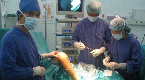
- These surgeries are all performed arthroscopically, meaning they are performed using a minimally invasive method. Arthroscopic surgery is performed through several small incisions to access the damaged portion of the meniscus. Images of the inside of the joint will be projected onto a screen, allowing the orthopedic surgeon to determine the location of the tear. From there, special instruments will be inserted to repair or remove damaged tissue.
4.3 Recovery
The time it takes for knee function to recover after a medial meniscus tear depends on the type of tear, the severity of the tear, and the treatments used to repair the damaged tissue.
In particular, if only non-surgical treatment is needed, the recovery process can take from six to eight weeks. In contrast, if corrective surgery is needed, such as in a grade 2 meniscus tear, the recovery process can take up to three months for repair surgery while only about three to four weeks for meniscectomy surgery.
In summary, the medial meniscus is located in the middle or inner part of the knee, acting as a shock absorber and providing stability to the knee. Although there are many degrees of medial meniscus tears, grade 2 medial meniscus tears are usually severe and require surgical intervention. After that, the patient needs to arrange a reasonable rest time combined with physical therapy exercises in stages to quickly restore knee function.
To arrange an appointment, please call … or make your reservation directly HERE. You may also download the MyVinmec app to schedule appointments faster and manage your reservations more conveniently.