Article written by: Microbiology Specialists, Laboratory Department, Vinmec Central Park International General Hospital
Candida albicans is a yeast that commonly causes infections in various parts of the human body, including the lungs. Accordingly, Candida can cause many dangerous complications in the lungs, so patients need timely diagnosis and treatment.
1. What Is Candida Albicans?
Candida albicans is a yeast fungus that commonly causes infections in various parts of the human body, including the lungs. Candida species are widely present in the environment and often reside naturally on the skin, mouth, gastrointestinal tract, and genital area without causing harm. Approximately 15–30% of healthy individuals carry Candida in the mouth and throat, and around 15% in the bronchi.
However, under favorable conditions, Candida can grow excessively and cause infections in other parts of the body.

2. Why Does Candida Infect the Lungs?
The isolation of Candida species from the respiratory tract is common in patients admitted to the ICU who require intubation or chronic tracheostomy. This generally reflects airway colonization rather than infection. Candida pneumonia and lung abscesses are rare, with only a few documented cases of primary pneumonia or abscess following aspiration of oropharyngeal secretions.
Candida pneumonia mainly occurs in patients with severe immunosuppression when the infection spreads to the lungs via the bloodstream. Chest CT scans typically reveal multiple lung nodules.
Pulmonary candidiasis: Candida albicans is one of the most common Candida species causing disease in immunocompromised patients or those with prolonged antibiotic nebulization. Fungal pneumonia is relatively uncommon but accounts for a small portion of pneumonia cases. Candida is the most common pathogen causing invasive fungal infections, representing 70–90% of cases.
Bronchial Candida infections: More frequently observed in children, where fungi from the mouth or throat are aspirated into the bronchi.
Trắc nghiệm: Làm thế nào để có một lá phổi khỏe mạnh?
Để nhận biết phổi của bạn có thật sự khỏe mạnh hay không và làm cách nào để có một lá phổi khỏe mạnh, bạn có thể thực hiện bài trắc nghiệm sau đây.3. Clinical Symptoms of Candida Lung Infection
The clinical symptoms of pulmonary Candida infections include:
Fever and cough (usually without sputum).
Pleuritic chest pain or discomfort.
Progressive shortness of breath leading to respiratory failure.
Symptoms of airway obstruction and mediastinal lymph node compression in endemic fungal diseases.
Lung consolidation and pleural friction rub
Localized Candida in the lungs may present as acute, subacute, or chronic disease:
Acute cases resemble bacterial pneumonia with symptoms like:
High fever (up to 39°C).
Paroxysmal cough producing gray or yellow sputum.
Hemoptysis (coughing up blood) and possible respiratory failure.
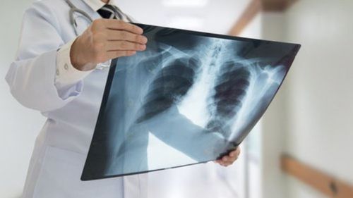
Chest X-rays reveal pulmonary infiltrates similar to pneumonia, affecting one lung (localized) or both lungs, resembling bronchopneumonia.
Bronchial Candida infections:
Common in children due to aspiration of Candida from the mouth or throat.
Symptoms include chest pain, bloody sputum, and wheezing similar to bronchial asthma due to an allergic response to Candida.
Bronchoscopy shows scattered white plaques, pseudomembranes, and inflamed bronchial mucosa.
4. Diagnosis of Pulmonary Candida Infection
4.1 Blood Tests
White blood cells (WBCs): Elevated in otherwise healthy individuals with endemic fungal infections.
Eosinophilia: Suggests an increased likelihood of opportunistic fungal infections like Candida or Aspergillus.
4.2 Gram Stain and Microscopy of Sputum
Proper transport, handling, and culturing of respiratory specimens are essential.
Detects fungal hyphae or yeast cells.
4.3 Culture and Identification of Candida Species
Respiratory samples, especially from bronchoalveolar lavage (BAL) or sputum, are cultured to isolate and identify Candida species.
Blood cultures confirm disseminated Candida infections.
4.4 Confirmatory Diagnosis
Histopathological examination of invasive infection is definitive.
Bronchial biopsy or transbronchial biopsy during bronchoscopy provides high diagnostic value.
4.5 Chest X-Ray
4.6 Bronchoscopy
5. Treatment of Pulmonary Candida Infections
Candida infections are caused by Candida yeasts, predominantly Candida albicans.
5.1 Treatment for Isolated Candida in Respiratory Secretions
Recommendation:
The presence of Candida in respiratory secretions often reflects overgrowth and rarely requires antifungal therapy (strong recommendation; moderate-quality evidence).
Summary of Evidence:
Although the diagnosis of Candida pneumonia is supported by isolating the organism from bronchoalveolar lavage (BAL) specimens, a definitive diagnosis requires histopathological evidence of invasive disease.
Clinical studies have shown that Candida isolated from BAL or sputum has low predictive value for invasive pulmonary candidiasis.
In a prospective study, none of the 77 patients who died in an ICU with clinical and radiographic evidence of pneumonia and positive Candida cultures from BAL or sputum demonstrated evidence of Candida pneumonia on autopsy. Due to the rarity of Candida pneumonia, the extremely common finding of Candida in respiratory secretions, and the lack of specificity of this finding, the decision to initiate antifungal treatment should not be made based solely on respiratory culture results.
Recent observations suggest that airway invasion by Candida species is associated with bacterial development and pneumonia. Invasive Candida in the airways is also linked to worse clinical outcomes and higher mortality rates in these studies. However, it remains unclear whether the isolation of Candida in the airways has a causal relationship with poorer outcomes or is merely a marker of disease severity.
5.2 Treatment of Oropharyngeal Candida Infections
Recommendations
Mild Infections:
Clotrimazole, 10 mg lozenges, 5 times daily for 7–14 days (strong recommendation; high-quality evidence).
Miconazole, 50 mg buccal tablets, applied once daily for 7–14 days.
Alternative Treatments:
Nystatin suspension (100,000 U/mL), 4–6 mL, 4 times daily for 7–14 days.
Nystatin lozenges (200,000 U), 4 times daily.
Moderate to Severe Infections:
Fluconazole, 100–200 mg orally daily for 7–14 days.
For Fluconazole-Resistant Cases:
Itraconazole solution, 200 mg/day.
Posaconazole suspension, 400 mg twice daily for 3 days, then 400 mg/day for up to 28 days.
Alternative options for fluconazole intolerance include voriconazole, 200 mg twice daily, OR oral AmB deoxycholate suspension, 100 mg/mL four times daily (strong recommendation; moderate-quality evidence).
Intravenous echinocandin (caspofungin: loading dose of 70 mg, then 50 mg/day; micafungin: 100 mg/day; or anidulafungin: loading dose of 200 mg, then 100 mg/day) OR intravenous AmB deoxycholate at 0.3 mg/kg/day are alternative options for intolerance (weak recommendation; moderate-quality evidence).
Chronic Suppressive Therapy:
Long-term fluconazole (100 mg, 3 times weekly) may be needed for recurrent infections.
HIV-Related Candida Infections:
Antiretroviral therapy (ART) is strongly recommended to reduce recurrence rates.
Candida Associated with Dentures:
Proper sterilization of dentures is necessary alongside antifungal treatment.
Summary of Evidence
Oropharyngeal and Esophageal Candida infections often occur in association with HIV infection, diabetes, leukemia, malignancies, steroid use, radiation therapy, antibacterial treatment, and denture use. These infections are recognized as indicators of immune dysfunction.
In HIV patients, oropharyngeal candidiasis is most frequently observed when the CD4 count drops below 200 cells/mL.
The advent of effective antiretroviral therapy (ART) has significantly reduced the incidence of oropharyngeal candidiasis and refractory cases.
Fluconazole resistance or multi-azole resistance typically results from prolonged and repeated exposure to fluconazole or other azole antifungals. In patients with advanced immunosuppression and low CD4 counts, resistance to C. albicans and the emergence of non-albicans Candida species (e.g., C. glabrata) have been documented, causing refractory mucosal Candida infections.
Most cases of oropharyngeal candidiasis are caused by C. albicans, either as a single infection or part of a mixed infection.
Symptomatic infections caused by C. glabrata, C. dubliniensis, and C. krusei have also been described.
Randomized prospective studies have been conducted on oropharyngeal candidiasis in AIDS and cancer patients. Key findings include:
Topical therapy is initially effective for most patients.
In HIV patients, symptoms often recur earlier and more frequently with topical treatments compared to systemic fluconazole.
A multicenter randomized study found that miconazole 50 mg mucoadhesive buccal tablets (once daily) were equally effective as clotrimazole 10 mg lozenges (five times daily).
Fluconazole and itraconazole solution demonstrated superior efficacy compared to ketoconazole and itraconazole capsules.
Posaconazole solution is as effective as fluconazole in AIDS patients.
Posaconazole delayed-release tablets (300 mg once daily) are approved for prophylaxis in high-risk patients, with stable bioavailability (~55%).
However, its efficacy for mucosal Candida infections remains under evaluation.
Recurrent infections often occur in patients with prolonged immunosuppression, especially those with AIDS and low CD4 counts (<50 cells/μL). Long-term suppressive therapy with fluconazole has been shown to be effective in preventing oropharyngeal candidiasis.
In a large multicenter study involving HIV patients, long-term suppressive therapy with fluconazole was compared to intermittent use of fluconazole in response to symptomatic outbreaks.
Continuous suppressive therapy was more effective in reducing the recurrence rate compared to intermittent therapy. However, continuous treatment was associated with an increased risk of in vitro drug resistance. The frequency of disease recurrence remained similar between the two groups.
Oral AmB deoxycholate, nystatin suspension, and itraconazole capsules are less effective than fluconazole in preventing oropharyngeal candidiasis. For infections that are fluconazole-refractory, initial treatment with itraconazole solution is recommended, with 64% to 80% of patients responding to this therapy.
Posaconazole suspension has shown efficacy in approximately 75% of patients with recurrent oropharyngeal or esophageal Candida infections, and voriconazole is also effective for fluconazole-refractory infections.
Intravenous caspofungin, micafungin, and anidulafungin have proven to be effective alternatives for patients who do not tolerate azole antifungals.
Topical or intravenous AmB deoxycholate has also shown effectiveness in some patients; however, pharmacists must prepare it as an oral formulation.
Immunotherapy with granulocyte-macrophage colony-stimulating factor (GM-CSF), macrophage activators, or interferon is occasionally used in the management of refractory oropharyngeal and esophageal Candida infections that are unresponsive to standard antifungal treatments.
Reduction in oral Candida colonization and a lower frequency of symptomatic oropharyngeal candidiasis have been observed in HIV patients receiving effective antiretroviral therapy (ART). Therefore, antiretroviral therapy should be initiated whenever possible for HIV patients with oropharyngeal or esophageal Candida infections.
Vinmec International General Hospital is a trusted medical facility for the examination, prevention, and treatment of various respiratory diseases, including pneumonia. With a team of highly qualified and experienced doctors, advanced equipment, and excellent medical services, Vinmec provides a comprehensive care process to ensure effective treatment and minimize complications caused by pneumonia.
For more health, nutrition, and beauty tips, visit Vinmec International Hospital to safeguard the health of yourself and your loved ones.
To arrange an appointment, please call HOTLINE or make your reservation directly HERE. You may also download the MyVinmec app to schedule appointments faster and manage your reservations more conveniently.


