The article is professionally consulted by Nguyễn Quang Đức, MSc, MD, Nuclear Medicine Doctor - Department of Diagnostic Imaging and Nuclear Medicine - Vinmec Times City International General Hospital. The doctor has over 12 years of experience in the field of Diagnostic Imaging.
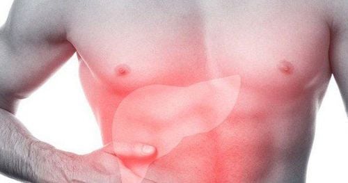
Liver ultrasound is a common imaging technique that uses ultrasound waves through a special probe placed on the patient's abdomen to observe the liver in black and white images. A liver ultrasound can help doctors detect abnormalities in the liver. So, what does it indicate when the liver ultrasound shows a hypoechoic mass?
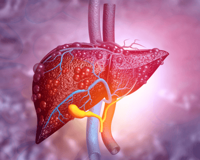
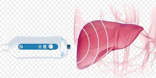
1. What is liver ultrasound?
Liver ultrasound is an imaging diagnostic method used to detect liver lesions that a patient may be experiencing.
It can detect liver damage, evaluate fatty liver condition, identify abscesses, parasites, liver cysts, liver tumors, and assess the severity of the disease, thereby determining an appropriate treatment plan.

2. What Can Liver Ultrasound Results Indicate?
- Liver ultrasound helps diagnose abnormal liver conditions and can be done alone or in combination with a general abdominal ultrasound. Some diseases detectable through liver ultrasound include hepatitis, cirrhosis, liver cancer, fatty liver, etc.
- For patients with hepatitis, liver ultrasound images show lesions in the liver parenchyma, and the liver may appear enlarged.
In patients with fatty liver, ultrasound images show increased brightness in parts or the whole liver, suggesting fat accumulation within liver cells. - In cases of cirrhosis, liver ultrasound shows changes in size and abnormalities in the liver parenchyma. In the early stage of cirrhosis, the liver may be enlarged, but as cirrhosis progresses, the liver shrinks and small nodules (under 1 cm) appear.
- Additionally, liver ultrasound can detect complications of cirrhosis, such as ascites, varicose veins, and splenomegaly, which are dangerous complications.
- For liver cancer, liver ultrasound plays a very important role in detecting abnormal masses, guiding early diagnosis and treatment.
- Besides common liver diseases, ultrasound can also help diagnose liver abscesses, liver fluke infections, liver cysts, and benign liver tumors.
3. Significance of a Hypoechoic Mass in Liver Ultrasound Results
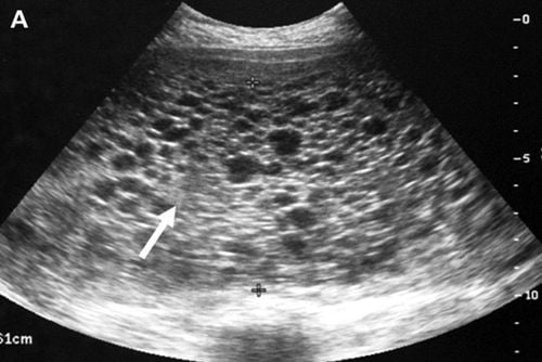
- Liver ultrasound results often describe whether there is a hypoechoic mass in the liver. If the result shows a hypoechoic mass or nodule in the liver, it may suggest a liver tumor.
- However, the presence of a hypoechoic mass in liver ultrasound cannot definitively diagnose whether the tumor is benign or malignant.
- The hypoechoic mass in liver ultrasound is valuable in providing preliminary information to guide doctors in treatment planning. It should be combined with other factors such as symptoms, clinical examination, medical history, and other tests (such as abdominal CT scan, liver cancer markers, etc.) to accurately diagnose whether the mass is benign or malignant.
4. Characteristics of liver tumors
Liver tumors, like other tumors, include two types: benign and malignant. Malignant liver tumors are more dangerous than benign ones, and most patients with advanced stages of malignant tumors can die quickly.
Therefore, early detection of the disease is crucial for timely treatment to avoid dangerous complications.
4.1. Benign Liver Tumors
- Some common benign liver tumors include hemangiomas, adenomas, benign nodular hyperplasia, liver cysts, etc. Most benign tumors develop slowly, do not have obvious symptoms, and do not cause discomfort for the patient.
- They are often discovered incidentally through a routine abdominal ultrasound, showing a hypoechoic mass in the liver, with cancer markers being negative.
- Benign liver tumors usually do not pose a threat to patients, so the treatment for benign liver tumors primarily involves a balanced lifestyle and regular monitoring to detect any abnormal signs of the tumor.
- In a small number of cases, intervention may be needed if complications such as rupture or bleeding within the tumor occur.
4.2. Malignant Liver Tumors
- Malignant liver tumors, also known as primary liver cancer, are dangerous diseases that can threaten the patient's life if detected in the late stages. Patients with liver cancer often experience symptoms such as pain in the upper right abdomen, jaundice, fatigue, weight loss, poor appetite, etc.
- In some cases, a small tumor may be detected in the upper right abdomen. A hypoechoic mass in liver ultrasound is considered an early indication before further tests are done to confirm the diagnosis of liver cancer.
- There are many causes of liver cancer, with the most common being chronic hepatitis B, hepatitis C, liver damage due to alcohol abuse leading to cirrhosis, and the development of malignant liver tumors.
- In addition, liver cancer can also manifest as a congenital form called Fibrolamellar carcinoma (also known as fibrous plate liver cell cancer).
- Primary liver cancer is a condition that can cause rapid death if detected late. Therefore, to reduce the risk of this danger, early detection through liver ultrasound showing a hypoechoic mass and positive liver cancer markers is crucial.

However, timely treatment protocols can offer hope for the patient. The treatment process for liver cancer depends on the stage and condition of each patient.
5. Notes Before Performing Liver Ultrasound
Some notes before performing liver ultrasound:
- About a week before the liver ultrasound, patients should avoid consuming foods that are difficult to digest or high in fat. Instead, opt for light, non-fatty foods to avoid putting pressure on the liver.
- It is recommended to perform liver ultrasound in the morning after fasting to ensure the most accurate results.
To arrange an appointment, please call … or make your reservation directly HERE. You may also download the MyVinmec app to schedule appointments faster and manage your reservations more conveniently.