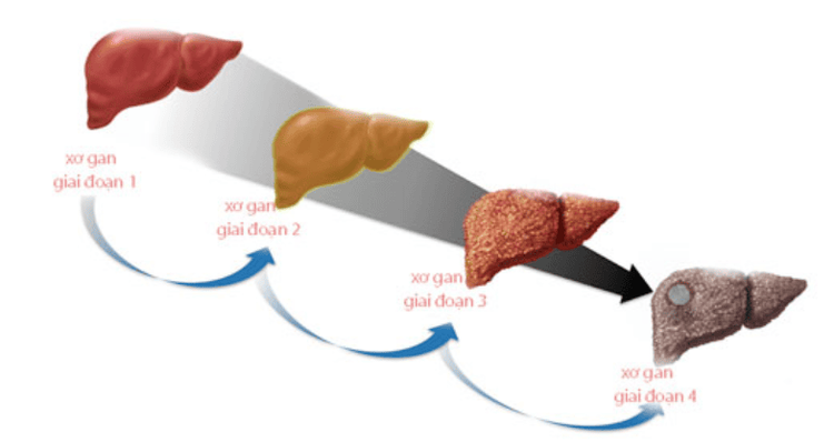
Liver fibrosis stages F2 and F3 represent the next progression of stage F1. These stages are considered the transitional phase between early-stage fibrosis and advanced liver cirrhosis (F4). At this point, fibrous and scar tissues have become more evident, with notable damage to liver function.
1. What is liver fibrosis F2-F3?
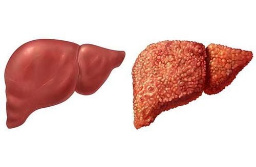
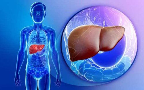
Liver fibrosis occurs when liver cells are damaged continuously over a long period, characterized by the replacement of liver tissue with fibrous tissue, scars, and regenerative nodules. This leads to impaired liver function.
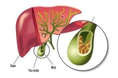
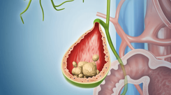
Causes of liver fibrosis include: Viral hepatitis (e.g., hepatitis B, C) ; Alcohol, smoking; Biliary diseases (e.g., bile duct obstruction, gallstones); Malnutrition; Obesity, diabetes; Parasitic infections (e.g., schistosomiasis, liver fluke); Drug and chemical toxicity; Primary biliary cirrhosis; Heart diseases, metabolic disorders and unknown causes.
Liver fibrosis F2 – F3 is the level and stage of the disease.
Liver fibrosis F2: The liver appears more damaged and has more symptoms.
This stage indicates more significant liver damage with increasing fibrous tissue. Fibrotic and scar tissues become more apparent. Excess connective tissue replaces normal liver cells, leading to reduced liver function, toxin accumulation, and metabolic disruptions.
Liver fibrosis F3:
At this stage, liver function is significantly impaired due to the predominance of fibrotic tissues over healthy liver cells. The remaining normal liver cells are overburdened, resulting in progressive damage and toxin buildup, which further accelerates the fibrosis process.
2. Can liver fibrosis F2-F3 be treated?
Liver fibrosis progresses through multiple stages, classified as F1, F2, F3, and F4.
- F1 and F2: These stages are considered mild to moderate and are potentially reversible if detected early and treated appropriately.
- F3 and F4: These stages represent severe fibrosis or cirrhosis. While complete recovery is unlikely, treatment focuses on symptom management, slowing disease progression, and extending the patient’s life expectancy.
3. Symptoms of liver fibrosis F2-F3
Symptoms of stage F2:
- Bloating, indigestion, and fatigue
- Mild fever, especially in the evenings
- Dark yellow urine, yellowish discoloration of hands and feet
- Intermittent pain in the right upper abdomen
- Nosebleeds, gum bleeding
- Dry and discolored nails
Symptoms of stage F3:
- Severe digestive issues, black stool, nausea, or vomiting blood
- Edema in the feet (indentation from pressure takes 1–2 minutes to disappear)
- Abdominal swelling due to ascites, accompanied by severe pain upon palpation
- Jaundice spreading from hands and feet to the entire body
- Increased heart rate, frequent dizziness, and fainting
Early detection of these symptoms is crucial for timely diagnosis and treatment.

4. Diagnostic methods for liver fibrosis
Blood tests:
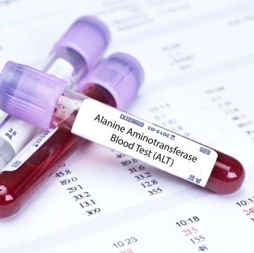
Fibrotic tissue in the liver obstructs normal blood flow, causing blood to back up into the spleen, leading to splenomegaly. The spleen then destroys red blood cells, white blood cells, and platelets, resulting in reduced blood cell counts (a condition known as hypersplenism). Low platelet levels on blood tests may indicate liver fibrosis.
Ultrasound:
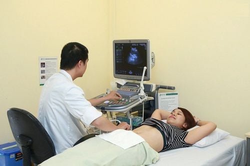
This non-invasive and widely available imaging method allows for the evaluation of liver size, structure, surface irregularities, and abnormalities like fatty liver, abscesses, or tumors. Doppler ultrasound can provide detailed views of liver blood vessels and flow directions.
Liver elastography (FibroScan):
This advanced diagnostic method assesses liver stiffness and fibrosis severity. During the procedure:
- The patient lies supine with their right arm positioned behind their head.
- The operator applies a probe to the intercostal area where liver biopsy is typically performed, applying light pressure.
- Ten consecutive measurements are taken at the same site, and the machine calculates an average stiffness value.
This method is non-invasive, suitable for patients with obesity or ascites, and applicable to various organs beyond the liver.
To arrange an appointment, please call HOTLINE or make your reservation directly HERE. You may also download the MyVinmec app to schedule appointments faster and manage your reservations more conveniently.

