This is an automatically translated article.
The article is professionally consulted by Master, Doctor Le Xuan Thiep - Department of Diagnostic Imaging - Vinmec Ha Long International Hospital
Currently, kidney transplantation has become a common technique for patients with end stage kidney disease. Ultrasound is a safe imaging method, plays an important role in the evaluation of patients after kidney transplantation, can detect anatomical, functional and vascular complications.
1. Transplant kidney ultrasound
A kidney transplant is a treatment for patients with end-stage kidney disease. A kidney transplant is the removal of a kidney from a healthy person or a healthy kidney from a brain-dead person to be transplanted into the abdomen, leaving the patient's old kidney intact. The most favorable place to place the new kidney is the right iliac fossa, there are also some cases on the left side. The ipsilateral iliac artery and vein will be connected to the renal artery and vein of the transplanted kidney, and the transplanted renal ureter will be sutured to the bladder. People only remove 1 or 2 pathological kidneys in some special cases such as: kidney disease with severe chronic inflammation, polycystic kidney too large, severe renal artery stenosis,... A patient can be transplanted. kidneys are many times, if the transplanted kidney is damaged.Renal ultrasound uses ultrasound waves with high frequencies beyond the hearing range of the human ear to create images of the size and structure of the kidney. From there, it is possible to evaluate the signs of kidney disease, or complications after kidney transplantation.
Ultrasound is a safe, non-invasive imaging method that can be performed in the ward and is widely used in the evaluation of patients after kidney transplantation. Ultrasound can detect complications after kidney transplantation including anatomical, functional, and vascular complications.

Siêu âm là một phương pháp chẩn đoán hình ảnh an toàn không xâm lấn, có thể thực hiện được ngay tại buồng bệnh và được sử dụng rộng rãi trong việc đánh giá bệnh nhân sau ghép thận
2. Complications after kidney transplant
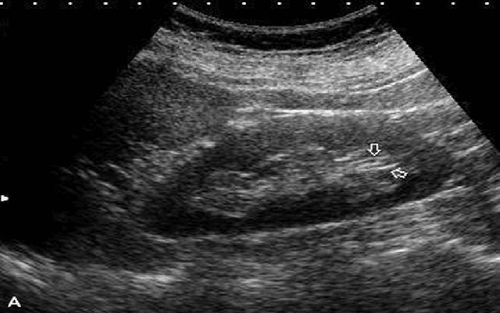
2.1 Anatomical complications Anatomical complications include perirenal fluid, hydronephrosis and lesions in the renal parenchyma.Perirenal fluid: Includes hematoma, urine, lymph cysts and abscesses. These are complications that occur in less than 50% of kidney transplant cases. Ultrasound shows a perirenal fluid mass, depending on the location and size of the fluid mass, it will affect the transplanted kidney, ureter or perirenal structures. Consequences can cause fluid retention in the kidneys, torsion, swelling of the legs, abdominal wall and genitals. Hematomas usually appear in the postoperative period within the first two weeks after surgery. The site of hematoma is usually in the subcutaneous tissue or around the transplanted kidney. The ultrasound image depends on the stage of the disease, with acute hematoma there will be an image of an empty volume of fluid, in the late stage there will be an image of a mixed-negative mass that may have an internal wall. Urinary cyst: is a sac of urine adjacent to the kidney that connects to the urinary tract. This is a serious complication, appearing in the first 2 weeks after surgery. The sonographic image is a homogeneous anechoic mass unless there is infection or internal blood. Lymphoid cyst: A later complication occurring 4 to 8 weeks after kidney transplantation. The site is usually between the bladder and the transplanted kidney. Lymphatic cysts are caused by disruption of the surrounding lymphatic channels. Ultrasound imaging reveals a mass with a clear or slightly concave cystic structure, mostly with an internal wall. Perirenal Abces: A rare complication that can occur in the early post-transplant period due to inflammation or infection of a urinary cyst, lymph node, or hematoma. Ultrasound imaging may show an echogenic or mixed perirenal mass. Hydronephrosis: Due to compression from the outside or due to kidney stones, anastomosis edema, blood clots and narrowing of the ureter. Localized lesions: Appearing in the parenchyma of the transplanted kidney may be hypoechoic, hyperechoic, or mixed. Causes may be nephritis, focal pyelonephritis, renal infarction, abscesses.

Biến chứng về giải phẫu bao gồm dịch quanh thận, thận ứ nước và các tổn thương trong nhu mô thận
Acute kidney transplant rejection occurs a few days after transplantation and peaks around 1-3 weeks post-transplant. Ultrasound images show that the transplanted kidney becomes edematous, spherical, the whole kidney is hypoechoic, poorly differentiated cortical marrow, and has an elevated RI. Chronic kidney transplant rejection begins 3 months after transplantation. Ultrasound shows that the transplanted kidney has thin parenchyma, mild hydronephrosis, echogenic renal sinuses, parenchymal calcification, hypoperfusion, normal or increased RI. Acute tubular necrosis: more common than renal transplant rejection, less conformational changes in renal parenchyma. Acute tubular necrosis occurs in the early transplant period shortly after the consequences of ischemia. Ultrasound showed an elevated RI. 2.3 Vascular complications Vascular complications include early complications of thrombosis in the renal arteries and veins, late complications of arterial and venous stenosis, post-biopsy complications including arteriovenous catheterization - renal veins and aneurysms. Vascular complications in kidney transplantation occur in less than 10% of cases. These are curable complications and ultrasound plays an important role in identifying the vascular complications of kidney transplantation.
Renal artery thrombosis and renal vein thrombosis: These are dangerous complications, common in the early postoperative period, which can lead to rapid kidney failure. Arterial thrombosis is a rare early complication caused by severe rejection and acute tubular necrosis or surgical technique. Doppler ultrasound showed no renal artery and vein outflow. Renal vein thrombosis is more common, usually occurring between the third and sixth days after acute kidney transplant, possibly due to renal vein compression by fluid masses, poor surgical technique, etc. Ultrasound shows enlarged, hypoechoic kidneys, no Doppler signal in the renal veins, elevated RI of the renal artery, often with reversed diastolic flow. Renal artery stenosis: Usually occurs in the first 3 years after kidney transplant surgery. This is a common vascular complication, the site of narrowing 50% of cases appears anastomosis. Doppler ultrasonography showed a narrow site with color aliasing with peak systolic velocities above 200 cm/s. Doppler spectrum in renal parenchyma with waveforms with curvilinear peaks due to increase in peak time, decrease in RI. Renal vein stenosis: Usually due to compression from the outside by fluid masses or fibrosis of the perivenous space. Doppler ultrasound showed narrow local aliasing, three to four times increase in velocity. Arteriovenous fistula and renal aneurysm: A possible complication after percutaneous kidney biopsy. Most lesions are small and not dangerous. However, large shunts can lead to renal ischemia, rupture at the arteriovenous shunt, and aneurysm that can cause hematuria or bleeding around the aneurysm.

Các biến chứng về mạch máu trong ghép thận xảy ra dưới 10% các trường hợp
Vinmec International General Hospital is a high-quality medical facility in Vietnam with a team of highly qualified medical professionals, well-trained, domestic and foreign, and experienced.
A system of modern and advanced medical equipment, possessing many of the best machines in the world, helping to detect many difficult and dangerous diseases in a short time, supporting the diagnosis and treatment of doctors the most effective. The hospital space is designed according to 5-star hotel standards, giving patients comfort, friendliness and peace of mind.
If you have a need for consultation and examination at Vinmec Hospitals under the nationwide health system, please book an appointment on the website for service.
Please dial HOTLINE for more information or register for an appointment HERE. Download MyVinmec app to make appointments faster and to manage your bookings easily.