This is an automatically translated article.
The article is professionally consulted by Master, Doctor Nguyen Van Huong - Department of Diagnostic Imaging - Vinmec Danang International General Hospital.
Today, the number of people suffering from diseases related to the kidneys and urinary tract is increasing. For early detection of kidney-related diseases, ultrasound is an extremely effective method. Renal ultrasound is used to determine the location, shape, structure, size, and relationship of the kidney to other organs.
1. Overview of the kidney
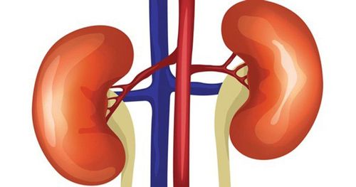
Kidneys are one of the special organs that play an important role in the urinary tract. The kidneys are located in the abdomen just behind the peritoneum connecting anteriorly to the abdominal aorta. Each person has 2 symmetrical kidneys on both sides of the lumbar spine across the thoracic vertebra from T1 to lumbar vertebra T3. At the upper pole and inward, the two kidneys are related to the adrenal glands, and at the lower pole, the two kidneys are related to the colon. In addition, the left kidney is related to the spleen and the right kidney is related to the liver.

2 quả thận đối xứng với nhau qua hai bên cột sống
2. What is renal ultrasound?
Ultrasound is a technique that uses ultrasound waves to create images of the size, structure, as well as pathological signs of many organs and internal organs of the body, including the kidneys. Renal ultrasound is used to determine the location, shape, size, and structure, and to assess the relationship between the kidney and other organs. Renal ultrasound is of high value in diagnosing kidney diseases such as: Kidney stones, kidney cysts, kidney abscesses, hydronephrosis, kidney tumors...
This is a non-invasive, painless method with low levels of pain. High accuracy and safety for the patient.
3. What diseases does renal ultrasound detect?
The ultrasound method often gives ambiguous results in the evaluation of diffuse kidney diseases such as nephrotic syndrome, glomerulonephritis (acute and chronic), renal starch infection. However, when these diseases have progressed to kidney failure and affect kidney size, ultrasound results are very clear. Because then the image of the kidney is smaller than normal, easy to recognize. Thus, in order to accurately diagnose diseases and disease progression, it is necessary to combine kidney - urological tests and multiple ultrasounds (if necessary) to help achieve high efficiency in treatment evaluation. .4. When is a kidney ultrasound needed?
Ultrasound is indicated in cases where the patient has signs of: Blood in urine, dysuria, urinary retention, kidney injury; renal, ureteral pain, renal failure (acute and chronic); Absence of renal shadow on radiographs, abrupt or elevated blood pressure in young adults, or suspected polycystic kidney disease.
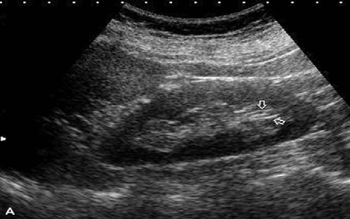
Khi có các dấu hiệu đau vùng thận bệnh nhân cần phải đi siêu âm để chẩn đoán
5. Signs and causes of small kidneys
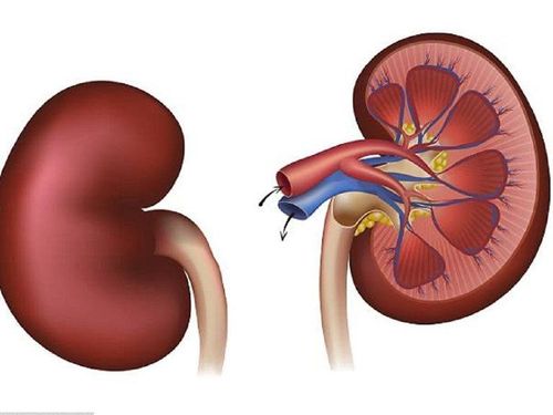
5.1 Normal kidney on ultrasound The kidney has an oval shape, the renal hilum is inside. Features of kidney size:● Varies by gender: men have larger kidney sizes than women.
● Varies with age: age-related decrease in kidney size also known as “age-related cortical thinning”, sometimes accompanied by increased fat in the renal sinuses.
● The size of the two kidneys is usually not the same, there may be a difference of 1-1.5cm.
● In adults, the right kidney is usually lower than the left kidney. Average height: 9-12cm; width 4-8cm; 3-5cm thick. If the kidney size is smaller than the above, it is called small kidney condition.
When the renal ultrasound is normal, we can see that the border is even, the left side has the spleen pressing on the base of the renal parenchyma with a triangular shape, in addition, the renal veins and arteries can also be clearly seen.
5.2 Causes of small kidney ● Congenital small kidney: People with congenital small kidney often have normal-looking kidneys but less than 5cm in height, compensatory enlargement of the symmetrical side kidney. The cause may be ischemic or total parenchymal scarring.
● Ischemia: narrowing of the renal artery leading to a decrease in kidney size. The ischemic kidney is usually small but has smooth margins and normal acoustical structure.
Post-traumatic renal atrophy: Most kidney injuries after a while will recover well without intervention, but still can leave scars. Renal atrophy is usually due to ischemia due to damage to the renal peduncle vessels, or due to the compression of chronic hematoma around the body, causing the kidney to tend to atrophy.
● Fatty kidney: is a disease related to the body's immune problems. Usually at the beginning of ultrasound, the kidney size is normal, the medullary cortical structure is still visible, but when the disease lasts for a long time, the kidney function gradually declines, causing more damage to the kidney tissue, the kidney will be small in size. gradually and in the final stage will lose the cortical structure.
● Chronic pyelonephritis: The main cause is chronic retrograde infection for a long time, causing fibrosis of the renal calyces, which will eventually lead to kidney failure and cause high blood pressure. The small kidney image on ultrasound is a partial reflection of the above phenomena, fibrous lesions can be seen in areas with thick ultrasound structure, kidney atrophy, and retracted calyces will be seen on ultrasonography. sound as small cysts in the periphery. However, these signs can also be seen in chronic glomerulonephritis.
Reflux nephropathy: is a disease that can lead to renal cortical scarring and reduced kidney size. Renal cortical scarring can be in one kidney or all of it. This case is called total scarring, the cortex is thin, the kidney size is reduced, the kidney image is small but the renal margin may be smooth.
Diabetes: can also be a disease that causes ultrasound to show small kidneys, hyperechoic kidney structures only appear at a late stage, which may be caused by vascular disease or infection.
In addition, there are many other causes of kidney size reduction such as: The degree of reduction in cortical changes (renal cortical necrosis, cortical thinning..), chronic pyelonephritis, gouty kidney disease, renal medullary cyst disease , kidney disease due to hypertension, late stage scleroderma, chronic lead poisoning..
Master Doctor Nguyen Van Huong used to be a Lecturer in Medical Imaging at Danang University of Medicine and Pharmacy, many years of experience working in hospitals such as: Cancer Hospital; Da Nang Hospital; Da Nang Obstetrics and Gynecology Hospital.
With his dedication to his work, dedication to the medical profession in general and the specialty of diagnostic imaging in particular, Dr. Huong is always loved and trusted by patients.
To register for examination and treatment at Vinmec International General Hospital, you can contact Vinmec Health System nationwide, or register online HERE.
Recommended video:
What should people with kidney stones eat?
SEE MORE
Children with nephrotic syndrome: Causes, symptoms, treatment Instructions on how to care & eat for people with kidney dysfunction What to eat with nephrotic syndrome?