This is an automatically translated article.
The article was advised by Assoc. Prof. TS.BS Tran Le Linh Phuong - Head of General Surgery Department, Vinmec Nha Trang International General Hospital.Chronic glomerulonephritis can progress to kidney failure, which is life-threatening if not detected early and with timely treatment measures. To know the exact condition of the kidney, the patient is forced to perform a kidney biopsy, read the structure of the kidney units under the microscope and combine many other tests.
1. How long does it take for chronic glomerulonephritis to progress to kidney failure?
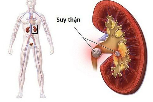
Renal failure is a condition in which kidney function declines, the kidneys cannot fully perform their functions, affecting the entire functioning of the body.
To know if chronic glomerulonephritis has progressed to kidney failure, for how long, patients need to perform tests to evaluate kidney function once a month, continuously for the first 3 months from at diagnosis and during treatment.
This is a chronic disease, so even when the disease is completely cured, the patient also needs to monitor and evaluate kidney function periodically every 3 - 6 months to check the status. If the disease is more stable, the time to perform renal function assessment can be extended to once a year.
If the results of the assessment of kidney function are good, the kidneys have not failed at the time of diagnosis and treatment, the disease responds to treatment, the patient has a reasonable lifestyle, and closely follows the doctor's instructions on With lifestyle, treatment, and completely negative proteinuria, chronic nephritis can prevent many types of chronic nephritis from progressing to kidney failure.
2. Tests to assess kidney function

Cần thực hiện một số xét nghiệm để đánh giá chức năng thận
To know the exact condition of the kidney, the patient is forced to perform a kidney biopsy, read the structure of the kidney units under the microscope and combine many other tests.
2.1. Biochemical tests Blood Urea Nitrogen and Creatinine are two substances that are eliminated from the body by the kidneys through urine. This is a product of the body's metabolism of protein. On average, Blood Urea Nitrogen: 6-24 mg/dL (equivalent to 2.5-8 mmol/L) and creatinine: 0.5-1.2mg/dL (equivalent to 45-110 mmol/L). Normal values may vary from laboratory to laboratory. If these values in the blood increase, it indicates that kidney function is worsening.
For more accurate results, the doctor may order the patient to perform additional tests of urea/blood and urea/urine, creatinine/blood and creatinine/urine. In normal subjects, creatinine clearance is 70-120 mL/min. If creatinine clearance is reduced, renal function is impaired.
2.2. Electrolytes Renal dysfunction causes an imbalance of electrolytes in the body, so one of the tests to evaluate kidney function should not be ignored is electrolytes.
Sodium (Sodium): In a normal person, blood sodium is 135 - 145 mmol/L. People with kidney failure can lose sodium through the skin, the digestive tract, or through the kidneys. Therefore, blood sodium in people with renal failure decreases. Manifestations of hyponatremia are mainly in the nervous system such as headache, nausea, vomiting, lethargy, more severe coma and convulsions. Potasium (potassium): In a normal person, blood potassium is in the range of 3.5 - 4.5 mmol/L. Renal failure causes a decrease in the excretion of potassium, causing an increase in blood potassium in patients with renal failure. Specific manifestations include: fatigue, paresthesias, gradual loss of reflexes, muscle paralysis, cardiac arrhythmia. Blood calcium: In a normal person, blood calcium is 2.2 - 2.6 mmol/L. People with kidney failure will have decreased blood calcium and increased phosphate. The main clinical symptoms are signs of neuromuscular stimulation such as muscle spasticity, increased tendon reflexes, convulsions, cardiac arrhythmias. 2.3. Acid-base balance disorder, normal blood pH at 7.37 - 7.43 allows optimal functioning of cell enzymes, muscle contraction proteins and clotting factors. Patients with kidney failure will have reduced excretion of acids formed during metabolism or loss of bicarbonate, causing metabolic acidosis in the body.
Acidosis causes respiratory disorders, cardiac arrhythmias, making hyperkalemia more serious.
Checking for acidosis is done by measuring blood pH or via bicarbonate.
Assess acidosis by measuring blood pH or indirectly with bicarbonate.
2.4. Blood uric acid Blood uric acid in normal people is as follows:
In women: 4.0 ± 1mg/dL (360 μmol/liter).
In men: 5.1 ± 1.0 mg/dL (420 μmol/liter).
Kidney damage causes uric acid in the blood to increase, especially in patients with kidney failure due to the inability to excrete uric acid from the body.
In addition to suggesting kidney failure, increased blood uric acid is also a manifestation of stones in the urinary system.
2.5. Urinalysis Total Urinalysis The urine density of an adult is normal in the range: 1.01 - 1.020. Decreased kidney function in the early stages will reduce the concentration of urine, causing the urine density to decrease accordingly. If renal failure is suspected, the patient will be ordered to do more tests to compare day and night urine density, urine concentration test, urine dilution test...
If the total urine sample is analyzed, If protein is present, the obtained results do not accurately assess the damage of the glomeruli, but it is suggestive for the patient to be indicated for a 24-hour quantitative proteinuria test.

Tổng phân tích nước tiểu và và máu để đánh giá tình trạng tổn thương của thận
2.6. 24-hour urine protein quantification Normal urine protein determination is from 0 to 0.2g/24h.
Proteinuria due to glomerular disease is usually persistent and greater than 0.3 g/l.
Diseases that can increase proteinuria such as: acute glomerulonephritis due to drug or chemical poisoning, kidney failure... or other diseases affecting the kidneys such as diabetes, lupus erythematosus, increased blood pressure...
2.7. Serum albumin Serum albumin in the normal range is about 35 - 50 g/L, accounting for about 50 - 60% of total protein. Acute glomerular disease causes a sharp drop in albumin.
2.8. Total protein in plasma The total protein in plasma reflects the filtering function of the glomerulus. Normally, the total protein in plasma is 60 - 80 g/L. Total protein is reduced when the glomerular membrane of the patient is damaged.
2.9. Complete blood cell analysis If a patient with renal failure has a decreased red blood cell count, he or she has chronic renal failure, especially when the decrease in red blood cell count is accompanied by no increase or decrease in reticulocytes. The patient may have iron deficiency anemia.
2.10. Abdominal Ultrasound Abdominal ultrasound can detect hydronephrosis due to ureteral obstruction. In case of bilateral hydronephrosis, it can cause chronic renal failure or acute renal failure.
In addition, abdominal ultrasound also helps to detect hereditary and congenital polycystic kidney diseases.
Through ultrasound images, if the kidneys are small in size or structurally abnormal, it may suggest chronic kidney disease.

Siêu âm bụng có thể phát hiện được tình trạng thận bị ứ nước do tắc nghẽn niệu quản
2.11. Abdominal CT scan Abdominal CT scan helps to clearly visualize the entire urinary system, thereby accurately detecting the disease. The method of imaging with contrast injection allows to reconstruct the urinary system image, not only detecting the disease but also clearly seeing the location and indicating the cause of ureteral obstruction.
Attention, only use CT scan in cases of suspected renal failure due to urinary tract obstruction.
2.12. Radioisotope renal scintigraphy Radioisotope renal scintigraphy is the only test that can evaluate the function of each kidney, clearly seeing the dialysis function, perfusion percentage, functional participation of each kidney.
Please dial HOTLINE for more information or register for an appointment HERE. Download MyVinmec app to make appointments faster and to manage your bookings easily.


