This is an automatically translated article.
The article is professionally consulted by Dr., Doctor Tran Nhu Tu and Master, Doctor Nguyen Thanh Nam - Department of Diagnostic Imaging - Vinmec Danang International HospitalSpongiform nephropathy, also known as spongiform nephropathy, is considered a benign disorder with a relatively low incidence. However, about 10% of patients have spongiform nephropathy, leading to the risk of diseases related to urinary tract infections and kidney stones, making patients struggle to filter kidney stones every year. Therefore, we should not be subjective and negligent in early detection and treatment of this disease.
1. What is spongy kidney disease?
Medullary sponge kidney (MSK), also known as Lenarduzzi Cacchi Ricci, is a congenital disorder of the kidney and is characterized by abnormal development of the renal pyramidal medulla, in which The small urinary ducts dilate to the cyst wall from 1 to 7 mm in one or both kidneys. These cysts are sponge-like, keeping urine flowing freely in the collecting ducts and kidneys. On ultrasound, the cysts can be seen and stones are easily seen in these cysts. Due to stagnant urine, people with spongiform nephropathy are more likely to develop kidney stones and urinary tract infections (UTIs) than the general population.
Bệnh dễ gây sỏi ở trong các nang ở 1 hoặc cả 2 bên thận
2. Symptoms of kidney spongiform disease
In its early stages, spongiform nephropathy may not present any specific symptoms. The first sign that a person with spongiform nephropathy may experience is a urinary tract infection or kidney stone. Both of these conditions have the same signs and symptoms such as:Burning or painful urination Pain in the back, lower abdomen or groin Unusual foul-smelling urine Foamy urine, dark or bloody The patient may have fever and chills Irritability, vomiting.
3. Causes of kidney spongiform disease
The exact cause of spongiform nephropathy is still unknown, which is the cause of cyst formation in the urinary ducts. Although spongiform nephropathy can be present in children from birth, most cases are not genetic.Many theories suggest that dilatation of the urinary ducts can be caused by uric acid causing obstruction in the embryonic cells or by hypercalciuria. The incidence of spongiform nephropathy is 1:5000 in which there is a high probability that people with kidney calcium stones (about 12-20%) will develop spongiform nephropathy later.
There are many factors that increase the risk of spongiform nephropathy, such as:
Hemihypertrophy (Beckwith Wiedemann syndrome) Caroli disease, manifested by dilation of the bile ducts in the liver, Ehlers Danlos syndrome causing joint damage supple and stretchy, delicate skin. Congenital cirrhosis of the liver Chronic kidney disease There is a family member with kidney disease
4. How to diagnose spongiform kidney disease?
To diagnose spongiform nephropathy, doctors will gather information from common medical diagnostic measures such as:
Intravenous pyelonephritis: Helps detect any blockage in the passageway urine and bright clusters shown in cysts. Computed tomography (CT) scan: Shows dilated or elongated urinary ducts. Ultrasound: helps detect kidney stones and calcium stones in the kidney.

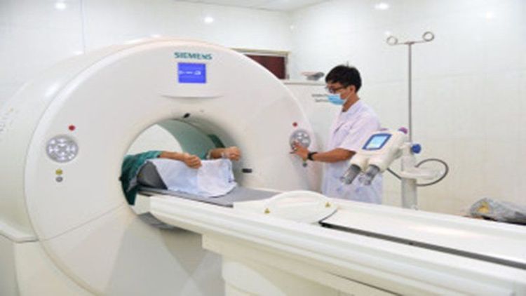
Chẩn đoán bệnh xốp thận bằng chụp cắt lớp vi tính
5. Ultrasound procedure of kidney spongiform disease
5.1. Ultrasound procedure of spongiform kidney disease Preparation for ultrasound
Before the procedure, the patient will be changed into specialized clothes and lying on the table, exposing the body part to be examined. The technician will apply a lubricating oil to the patient's skin to reduce friction when moving the transducer on the skin and support the propagation of sound waves. The transducer sends high-frequency sound waves through the patient's body. Sound waves will be partially blocked by bones or organs and reflected back to the computer. Depending on the area being examined, the patient may need to change positions in order for the technician to capture the best image. Perform ultrasound
Both kidneys will be scanned in multiple layers in the longitudinal and transverse planes to closely observe the entire renal volume. The patient usually has to lie on his side, the body is slightly turned inward to make the ultrasound process easier, recording the entire kidney
The right kidney will be scanned from the anterior lateral plane using the liver as an acoustic window. To view the lower pole of the kidney, scan posteriorly. The medical technician will ask the patient to take deep breaths to move the kidneys away from the ribs and intestinal gas. The left kidney is scanned posteriorly. The upper pole can be scanned through the spleen, but most of the time it will have to be scanned through the lumbar muscle causing the quality of the ultrasound image to be reduced.. After the ultrasound
After the ultrasound, the technician will wipe the gel off the patient's skin. . Total ultrasound time is usually less than 30 minutes, depending on the area being examined. The patient can resume normal activities immediately after the end of the ultrasound.
5.2. Ultrasound image of spongiform nephropathy In spongiform nephropathy, an ultrasound image of spongiform nephropathy will show a thick echogenic renal medulla with echogenic spots, some with a dorsal shadow. Ultrasonography will help distinguish spongiform nephropathy from papillary necrosis and polycystic kidney. To achieve the best diagnostic effect, it should be combined with contrast-enhanced nephrography.
Ideally, when there is suspicion and symptoms of urinary tract infection (difficulty urinating, burning urine, painful urination, fever, cloudy urine, it is advisable to go to the hospital for timely antibiotics. Pain or pain when urinating, suspected urinary tract infection, kidney stones should pay attention to look for more signs of kidney spongiform disease through medical techniques such as ultrasound, X-ray, computed tomography CT.
Currently, at Vinmec International General Hospital, there is a full range of modern equipment and facilities for early diagnosis.To register for examination and treatment at the Hospital, you can contact the System. Vinmec Health nationwide, or register online HERE
Recommended video:
What should people with kidney stones eat?
SEE MORE
Child nephrotic syndrome: Causes, symptoms, treatment Treatment Instructions on how to care & eat for people with kidney dysfunction What should eat well with nephrotic syndrome?


