This is an automatically translated article.
The article is professionally consulted by Master, Doctor Nguyen Thuc Vy - Doctor of Diagnostic Imaging - Department of Diagnostic Imaging - Vinmec Nha Trang International General Hospital.Kidney diseases can have serious effects on a person's health and are difficult to control. Applying renal ultrasound in the diagnosis and treatment of kidney disease is one of the most effective methods today.
1. What is renal ultrasound?
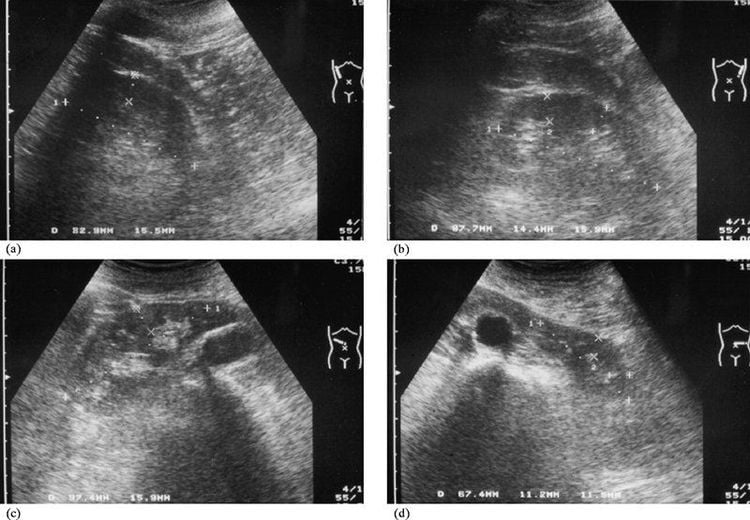
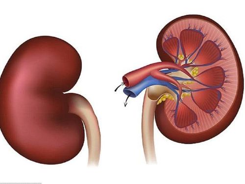
Kidney ultrasound, which is a modern, non-invasive technique, uses high-frequency sound waves to create realistic images of the structure, size, and typical signs of the disease. kidney related.This method is often used to determine the location, shape, size and structure of the kidney as well as the relationship between the kidney and other organs in the patient's body. In particular, renal ultrasound brings high value in the diagnosis of diseases located in the kidney such as: kidney stones, kidney failure, hydronephrosis, renal abscess.
Not only is it a non-invasive method, but this technique also minimizes pain for the patient, has a high degree of accuracy and ensures safety.
2. What diseases does kidney ultrasound help detect?
Renal ultrasound helps to detect diseases such as: acute glomerulonephritis, nephrotic syndrome, amyloidosis and chronic glomerulonephritis. If not detected in time, these diseases will progress to the stage of kidney failure and cause quite serious effects on the size of the kidneys, then performing an ultrasound examination of the kidneys will give clear results. .Therefore, in clinical diagnosis, doctors will assign patients to perform kidney-urinary tests, combined with renal ultrasound images to give the most accurate results, including: : urine tests, X-rays and blood tests to evaluate kidney function,...

3. In which case is renal ultrasound indicated?
A kidney ultrasound may be ordered periodically during general checkups of your entire body. If the patient appears abnormal signs related to the urinary tract, it is advisable to conduct a timely examination and perform uro-renal ultrasound.Below are unusual cases when a patient has kidney problems and will be assigned to perform a kidney ultrasound:
Blood in the urine. Painful urination, difficulty urinating, or the patient has urinary retention and is unable to urinate. Urine has abnormal symptoms such as: urine appears strange color such as cloudy urine, white color, urine with a lot of sediment, or urine with a lot of foam, ... Abdominal pain in the kidney area. The accident caused abdominal injuries. Sudden increase or decrease in blood pressure for no apparent reason. X-ray of the kidney area but no kidney is found or although the kidney is found, it is found in an abnormal position. Having kidney diseases: Polycystic kidney, kidney failure. Monitor and check kidney status. The images help diagnose ultrasound images of normal kidneys Specialist doctors will rely on the following renal ultrasound results to make an accurate diagnosis of the patient's health condition

4. Features showing that the ultrasound image of the kidney shows pathology
4.1 Diffuse pathology Features of this pathology include:Chronic glomerulonephritis : Ultrasound image shows small kidney size, irregular contours, and unclear medullary-cortical limits. Acute glomerulonephritis: When the size of the kidney is larger than normal, the contours are smooth and the medullary-cortical boundaries are clear. Renal tuberculosis: When the size change is not obvious, with irregular contour and in the patient's body, the image of water retention is seen in each area, there are calcified nodules appearing in each kidney. Total fluid retention due to ureteral stenosis in the patient with tuberculosis. Polycystic kidney disease: When the size of the kidney is large and the contour is irregular, and the medullary-cortical limit disappears, the whole kidney area appears many cysts, these cysts do not communicate with each other, and The patient may have polycystic hepatopancreas. 4.2 Focal kidney disease Kidney stones: When the ultrasound image of the kidney is dome-shaped (light-shaped) hyperechoic shadows, mostly found in the renal pelvis, or calyces, with calcified nodules in the renal parenchyma. of coral gravel will usually be many stones lying consecutively and in succession. Hydronephrosis: Renal ultrasound images provide an assessment of damage between the calyx, the renal pelvis, and the ureters. Perirenal hematoma: The image is a crescent-shaped fluid that surrounds the kidney, and can displace the kidney. Injury to the kidney: Visible images on renal ultrasound show characteristics of the extent of the injury. Kidney tumor: Usually ultrasound will identify the tumor but cannot distinguish between benign and malignant tumors. If you detect the typical symptoms of the diseases mentioned above, it is necessary to quickly find a reputable medical facility to conduct a kidney ultrasound in combination with the necessary tests to make the most accurate conclusion. .
Master. Doctor Nguyen Thuc Vy has 09 years of experience in Diagnostic Imaging. Dr. Vy has many years of experience working in the Department of Diagnostic Imaging at the University of Medicine and Pharmacy Hospital in Ho Chi Minh City, trained and attended many courses on specialized imaging at the University of Medicine and Pharmacy Hospital. Hue Pharmacy, Ho Chi Minh City University of Medicine and Pharmacy, Cho Ray Hospital. Currently working at the Diagnostic Imaging Department of Vinmec Nha Trang International General Hospital.
Please dial HOTLINE for more information or register for an appointment HERE. Download MyVinmec app to make appointments faster and to manage your bookings easily.


