This is an automatically translated article.
Article by Master, Doctor Mai Vien Phuong - Gastrointestinal endoscopist - Department of Medical Examination & Internal Medicine - Vinmec Central Park International General Hospital.
Before medical AI-assisted cytology becomes a common practice, significant barriers such as patient acceptance or regulatory issues need to be addressed careful. The reason is due to the development of CAD algorithms and artificial intelligence that can promote, shape and improve decision making in the management of colorectal lesions.
1.The role of AI-assisted cytoscope in colorectal cancer A major breakthrough in technological developments over the last decade enables real-time in vivo histology of the gastrointestinal tract only by pressing a button. Emerging cytoreductive-CAD offers the possibility of clearer delineation between benign and cancerous colon lesions. Furthermore, this new diagnostic tool contributes to the detailed detection and features of gastrointestinal tumors.
Thus, with emerging artificial intelligence in colonoscopy, treatment options for colonic lesions become more accessible and accurate.
Artificial intelligence technology provides "real-time" histology, thus determining whether a sizable colonic lesion (> 2 cm) should be treated with surgery or laparoscopic resection. Artificial intelligence endoscopy significantly shortens the final histological and endoscopic diagnosis of colonic lesions, avoiding unnecessary tissue biopsies.
2. What do the studies conclude? Lui et al. supported a study to evaluate the application of artificial intelligence-assisted image classifiers in medical care, to determine the feasibility of laparoscopic surgery for these patients. large colonic lesions based on unmagnified endoscopic images. They collected an artificial intelligence image classifier using 8000 endoscopic images of colonic lesions. Meanwhile, the validation set included 567 endoscopic images from 76 patients. The histological findings of resected specimens were used as a gold standard for validation in the study.
Therapeutic endoscopic resection is performed only in patients with well-differentiated adenocarcinoma with submucosal invasion ≤1 mm and without any lymph node involvement. The results obtained by the artificial intelligence image classifier are compared with those taken by (experienced) endoscopists. In patients with previously mentioned lesions, who were indicated for laparoscopic surgery, artificial intelligence had high accuracy (85.5%). This study highlights the clinical significance of artificial intelligence in predicting laparoscopic resection of large colonic lesions (>2 cm).
3. Artificial intelligence provides valuable information
According to Ichimasa, are the first to publish a paper on the role of artificial intelligence in predicting lymph node metastasis (LNM) in patients with invasive colorectal cancer (CRC). Their study aimed to show that artificial intelligence provides valuable information on the need for additional surgery after laparoscopic resection for invasive CRC pT1 colorectal cancers. .
One of the key endpoints in deciding on additional surgery in patients undergoing laparoscopic resection of invasive stage colorectal cancer is the presence of lymph node metastases. To minimize the need for additional surgery, the authors used an artificial intelligence model to predict the likelihood of lymph node metastasis in T1 CRC patients. The prediction data for lymph node metastasis was compared with the data of the Japanese, European and American guidelines.
4. The role of cytoscopy in the diagnosis of inflammatory bowel disease
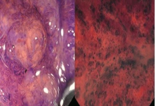
With the significance of endoscopic cytology-CAD in clinical practice, the diagnosis of patients with inflammatory bowel disease (IBD) has been significantly improved. This new endoscopic method enables real-time histological diagnosis and prediction of disease outcome. Bessho et al established the endoscopic cytoreductive score system (ECSS) to evaluate patients with IBD. The cytoscopy scale assesses the shape, spacing of the code segments, and visibility of the superficial microvessels.
Severity rating system according to histological changes of colonic mucosa. The authors also demonstrated a good correlation between Matt's scoring system and histological classification. The cytoscope score was graded by Ueda et al in 2018 by adding the index: Mucosal surface characteristics.
Another benefit of this upgraded point system is the ability to predict disease recurrence. Using probe-based cyto endoscopy with 1390x magnification, Neumann et al elucidated the role of cytoscope in the identification of mucosal cell structures in IBD patients. The system allows detailed analysis of ultrastructural samples such as nuclear-cytoplasmic ratio, size and shape of the nucleus. The collected data provide reliable differentiation of different types of inflammatory cells in the colonic mucosa.
In another study by Neumann et al., 100% concordance between the standard histopathological classification and cytoskeletal data was established. Another fascinating study by Nakazato et al., including 64 patients in clinical remission (Mayo score 0 and Geboes score ≤ 2), showed that the Cytoscopy Scale has high accuracy for remission. histology. In conclusion, they accept that the Cytoscopy Scale can be a reliable evaluation tool for the assessment of histological wounds.
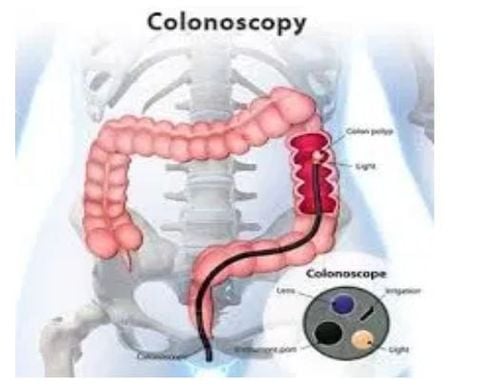
In their study, 187 patients with ulcerative colitis had white light endoscopy performed to determine the Mayo-endoscopic scoring of the colonic mucosa. After identifying the most severely inflamed area, they used cytoscope with the NBI regimen. Their analyzes showed that cytoscopy-CAD identified persistent histological inflammation with a sensitivity of 74% and a specificity of 97%. Maeda et al also showed that endoscopic cytology-CAD has added benefits for future therapeutic strategies. However, the authors considered conducting more studies because of the insufficient number of imaging studies.
6. Conclusion Before medical artificial intelligence-assisted cytoreoscopy became a common practice, significant barriers such as patient acceptance or implementation by an internist were present. Poor screening and regulatory issues need to be handled with care. The development of CAD algorithms and artificial intelligence can promote, shape, and improve decision making in the management of colorectal lesions. In general, cytoscopy has shown excellent accuracy, helping to support in vivo diagnosis of lesions in the lower gastrointestinal tract.
Please dial HOTLINE for more information or register for an appointment HERE. Download MyVinmec app to make appointments faster and to manage your bookings easily.
References:
1. Neumann H, Fuchs FS, Vieth M, Atreya R, Siebler J, Kiesslich R, Neurath MF. Review article: in vivo imaging by endocytoscopy. Aliment Pharmacol Ther . 2011; 33 :1183-1193. [PubMed] [DOI]
2. Sasajima K , Kudo SE, Inoue H, Takeuchi T, Kashida H, Hidaka E, Kawachi H, Sakashita M, Tanaka J, Shiokawa A. Real-time in vivo virtual histology of colorectal lesions when using the endocytoscopy system. Gastrointest Endosc. 2006; 63 1010-1017. [PubMed] [DOI]
3. Takamaru H , Wu SYS, Saito Y. Endocytoscopy: technology and clinical application in the lower gastrointestinal tract. Transl Gastroenterol Hepatol . 2020; 5:40. [PubMed] [DOI]
4. Inoue H , Kudo SE, Shiokawa A. Technology insight: Laser-scanning confocal microscopy and endocytoscopy for cellular observation of the gastrointestinal tract. Nat Clin Pract Gastroenterol Hepatol . 2005; 2:31-37. [PubMed] [DOI]


