This is an automatically translated article.
Article written by Dr. Nguyen Le Thao Tram - Diagnostic Imaging Doctor, Diagnostic Imaging Department - Vinmec Nha Trang International General Hospital
Non-mass lesions account for about 5% of lesions in breast ultrasound. These lesions are also called focal opacities, tubular structures, ductal changes, or non-massive lesions. Non-massive lesions have a fairly broad spectrum of pathology. This spectrum ranges from benign (fibrous changes or stromal fibrosis) to ductal carcinoma in situ to invasive cancer.
According to Korean breast radiologists from CHA Bundang Medical Center of CHA University in Gyunggi-do, knowledge and understanding of non-massive lesions are scarce. They investigated 119 women with non-massive lesions to determine their final outcome, and identified imaging features that differentiate malignancy from benign lesions.
In the Acta Radiologica article, the study analyzed 119 consecutive patients (21 to 69 years old) with 121 non-massive lesions identified on breast ultrasound over a 4-year period. 95 women of age were screened and graded mammograms.
The authors have defined non-massive lesions as those that are visible in two orthogonal planes, but cannot be described as a distinct mass because of indistinct margins or shape. Because the imaging characteristics of non-massive lesions on ultrasound have not been standardized, the authors classified based on distribution location and combined features. Non-massive lesions are classified into focal, line-lobular, or regional distribution. Features of non-massive lesions are also classified according to calcifications, structural disturbances, or ductal abnormalities.
A total of 33 malignancies in 33 patients were pathologically confirmed. The majority of malignancies (51.5%) were diagnosed as DCIS (ductal carcinoma in situ) followed by invasive ductal carcinoma (24.2%). 82 benign diseases were identified in 81 patients, with the majority being fibrous changes (28%), stromal fibrosis (25.6%), and fibroadenoma (14.6%).
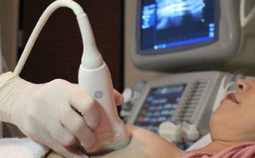
Siêu âm tuyến vú có thể phát hiện các tổn thương không khối
Lead author Jong Won Park MD and coauthors reported that 60% of melanoma patients had a palpable mass - the only symptom they reported - compared with 22% of patients with a symptomatic benign lesion. (Among women with benign lesions, only 9% reported a palpable mass).
Age and breast tissue density on mammography were not significantly different between patients with malignant and benign lesions. Calcification associated with asymmetry seen on mammography was more common in malignant lesions (37.5%) than in benign lesions (6.3%).
Breast ultrasound has shown a significant difference in the distribution between malignant and benign lesions. Linear or lobular distribution was found in 45.5% of malignant lesions compared with 17% of benign lesions. The local distribution was seen in 60.2% of benign lesions compared with 33.3% of malignant lesions. Calcification (27.3% vs 10.2%) and structural disturbances (18.2% vs 4.5%) were also more common in malignant lesions than in benign lesions. .
They conclude: “Non-massive lesions on breast ultrasound show a high risk of malignancy... Non-massive lesions must be managed based on clinical, mammographic, and ultrasonographic findings". They say additional research is needed, preferably by multiple institutions with larger research groups.
This article is based on a study by the authors Park Jong Won, Ko Kyung Hee, Kim Eun-Kyung, Kuzmial M Cherie, Jung Hae Kyoung, in 2017, “Non-mass breast lesions on ultrasound: final outcomes and predictors of malignancy.”, of Acta Radiol magazine.
Please dial HOTLINE for more information or register for an appointment HERE. Download MyVinmec app to make appointments faster and to manage your bookings easily.
The article references the source: appliedradiology.com