This is an automatically translated article.
The article was professionally consulted by Specialist Doctor I Le Hong Lien - Department of Obstetrics and Gynecology - Vinmec Central Park International General Hospital.Currently, breast cancer is trending younger. According to statistics of the International Agency for Research on Cancer (IARC) of the World Health Organization (WHO), breast cancer is the most common cancer in women. In Vietnam, in 2018, there were over 15,000 new cases, including more than 6,000 deaths from breast cancer. Mammography to identify breast calcification plays an important role in the prevention and early treatment of the disease.
1. What is mammary calcification?
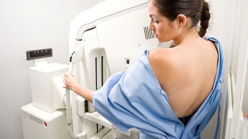
Benign breast calcifications identified by mammography are typically round, enlarged, and coarse with smooth margins and are more recognizable than malignant calcifications. Breast calcifications associated with melanoma (as well as many benign calcifications) are usually very small and require a magnifying glass to be seen more clearly.When it is not possible to give a specific etiology, the description of breast calcifications should include both their morphology and distribution. However, they should be reported if the physician reading the film is concerned that other film readers may misinterpret them.
2. What is breast ultrasound?
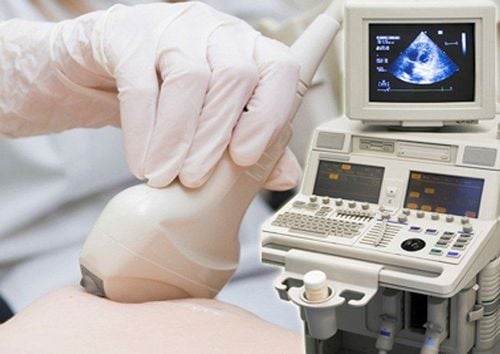
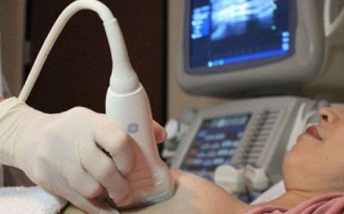
Mammography can be performed in the usual way by direct examination by a doctor or by 3D automatic breast ultrasound. With the 3D automatic breast ultrasound method, the patient will be examined by an automatic scanner. The specialist will then analyze the resulting images to look for abnormalities of the mammary glands.
The purpose of breast ultrasound is to detect lesions of the mammary glands. Breast ultrasound is being applied worldwide for diagnosis, early detection and monitoring of mammary gland abnormalities, especially breast cancer.
3. Benign breast tumor on ultrasound
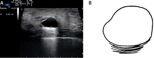
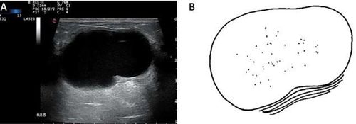
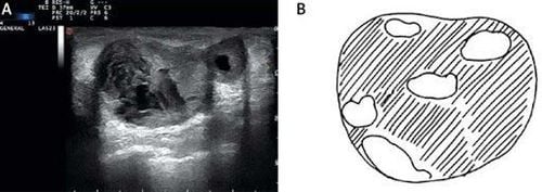
Many studies have shown that benign breast tumors often have the following signs on ultrasound:Clear, tender margins; Limitation of the lesion: The face is clearly and thinly separated from the adjacent healthy tissue (additional sign); Calcification: Coarse calcification; Direction: Parallel; Echoic structure: Drum (cystic) echo, thick echo, posterior tone change: hyperechoic, homonymous or slightly hypoechoic compared with mammary parenchyma; Has a thin shell; An ellipse in which the major axis is parallel to the skin surface; The block does not cause tension in neighboring structures; There are no signs suggestive of malignancy; Classification of benign breast tumors:
Type 1- Simple cyst: Round or oval cyst, clear margin, negative void, with increased illumination of the posterior wall;
4. Characteristics of malignant breast tumors
Shape: Irregular (main sign); Round. Direction: Not parallel (main sign); Shoreline: Tentacles (main sign); Unknown (additional sign); Corner flexion (additional sign); Minor polytone (minor sign). Lesions: Boundary unknown with thick echogenic margin (additional sign); Echoic structure: Very poor reverb (additional sign); Mixed reverb (additional sign). Change the tone behind: Shading the back of the back (additional sign); Calcification: Microcalcification (main sign); Doppler ultrasound; Intra-tumor perfusion; RI > 0.83, PI > 1.6. Metastatic lymph nodes: Short diameter > 1cm; Thickness of lymph node > 3mm; Loss of umbilical lymph nodes; Extra-umbilical lymph node perfusion; Multi-arc shore.5. Where is a good breast ultrasound?

The early stage breast cancer screening package 2020 will help a lot in the treatment process. Early intervention by methods such as using drugs, radiation therapy, chemotherapy, surgery, hormonal therapy, biology... can minimize the effects of the disease, slow down the progression of the disease, especially saving the lives of breast cancer patients.
Currently, on the Vinmec International General Hospital system, there are modern breast ultrasound machines to help diagnose early calcified nodules. Vinmec uses 3D breast ultrasound device Invenia ABUS - the most advanced automatic ultrasound device in the world today in breast cancer screening with many outstanding advantages compared to other ultrasound techniques:
The system is capable of screening, diagnosing, monitoring before, after, treating breast diseases and detecting breast cancer with accuracy up to 90%. In addition, the mammography system helps diagnose early breast calcifications, increasing the ability to detect breast cancer at a very early stage when there are no clinical manifestations; Integrating ARFI tissue elastic technology, assessing tissue stiffness helps verify and complement the results on 3D mammograms, increasing accuracy in the diagnostic process; The 3D breast ultrasound system helps to create anatomical sections that are currently not possible with traditional ultrasound systems; Detect and identify small lesions to support effective disease monitoring and treatment; There are data to assess breast lesions quickly, safely because it is non-invasive and non-radioactive, so pregnant women can use this system.
Please dial HOTLINE for more information or register for an appointment HERE. Download MyVinmec app to make appointments faster and to manage your bookings easily.


