This is an automatically translated article.
The article is professionally consulted by MSc, BS. Dang Manh Cuong - Radiologist - Radiology Department - Vinmec Central Park International General Hospital. The doctor has over 18 years of experience in the field of ultrasound - diagnostic imaging.Axillary lymph node ultrasound is a simple imaging method that helps doctors diagnose some diseases of axillary lymph nodes. This is also the first indicated method in cases of abnormality in axillary lymph nodes and suspected metastatic lesions in cancer pathology.
1. What is axillary lymph node ultrasound?
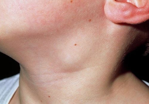
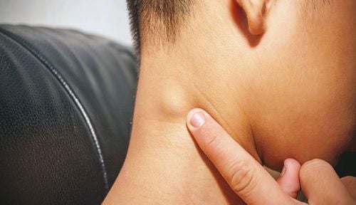
The axillary lymph node is one of the body's lymph nodes, involved in the process of eliminating or destroying disease-causing microorganisms in the body. Normally we rarely feel the axillary lymph nodes, but in some cases axillary lymph nodes are enlarged due to the body's immune response so we can feel them. The lymphadenopathy can be caused by many causes, so to determine part of the cause of axillary lymphadenopathy, we use the lymph node ultrasound method.Axillary lymph node ultrasound is a non-invasive, painless technique that uses an ultrasound machine with a flat transducer, high frequency to survey images of the axillary fossa to detect abnormal lymph nodes or pathological lesions. of the ganglia. Through the images obtained when performing an axillary lymph node ultrasound, the doctor can give directions for pathological diagnosis.
Sometimes axillary lymph nodes are distended due to a normal reaction after vaccination against tuberculosis. Ultrasound shows a solitary lymph node, usually round or oval in shape, hypoechoic compared with surrounding parenchyma, smooth border, chemical fluid...
In addition to the usual inflammatory reactions, lymphadenopathy can also be abnormalities that need to be diagnosed. Thanks to the ultrasound of the axillary lymph nodes, it is possible to detect the status of metastatic cancer in the lymph nodes, lymph nodes... through the signs suggested on the ultrasound image.

Hạch nách là một trong những hạch lympho của cơ thể, tham gia vào quá trình loại trừ hay tiêu diệt những vi sinh vật gây bệnh cho cơ thể
2. Indications and contraindications for axillary lymph node ultrasound
Indications for ultrasound of axillary lymph nodes:Suspicious lesions of cancer in organs that may metastasize to axillary nodes such as breast cancer, lymphoma, stomach cancer... Abnormally painful and swollen lymph nodes. Lymph node ultrasound guides the localization of cytological aspiration or histopathological biopsy to diagnose benign or malignant lymph node involvement. Contraindications: This is a non-invasive method, with no contraindications if there is no accompanying intervention. In case of ultrasound-guided cytology, it is contraindicated in cases of coagulopathy.
3. Steps to conduct axillary lymph node ultrasound
3.1 Preparation Performers include radiologists, technicians. Equipment: Ultrasonic machine with flat probe, frequency above 7.5MHz, gel used in ultrasound, cloth to wipe gel after ultrasound. Patients: No special preparation is required, the diagnostic ultrasound method is well explained to coordinate implementation.
Trước khi siêu âm, người bệnh sẽ được giải thích rõ về phương pháp siêu âm để phối hợp thực hiện
Steps to take:
Step 1: Anterior bilateral mammogram to find all abnormalities in all areas from areola to the periphery. Usually, during ultrasound, the transducer is moved in a circular, clockwise or counter-clockwise direction from the center of the mammary gland to the periphery. Step 2: The probe is brought to scan the axillary region, the slice is perpendicular to the humerus cap, allowing to clearly see the shoulder joint socket on the probe side and see the joint space, the contour around the joint socket of the humerus. , shoulderblade. Investigate lesions in this area. Step 3: Identify brachial vascular bundle on cross section with artery and vein. It is best to monitor blood vessels on color Doppler ultrasound. Step 4: Examine the lymph node system by rotating the transducer with a slice along the vascular bundle and moving downward parallel to the vascular bundle toward the anterior axillary line. Examining many damaged lymph nodes in many different locations, it is necessary to map out the exact position, depth and distance from the skin, large blood vessels to facilitate the clinician's guide to locate or puncture. cytology or histopathology. Step 5: Evaluate the weighing results describing the location, size, shape of the lesion, border, sheath of the lymph nodes, the amount and degree of perfusion... and other tests needed to make an accurate diagnosis. In order for the clinician to guide the diagnosis and next indication: Normal lymph node: Small in size, only a few millimeters, the shell is usually thin, the hilar node is deviated to one side with clear boundaries. Inflammatory-reactive lymph nodes: There are often many separate raised lymph nodes, small and similar in size, hypoechoic, well differentiated, oval or flattened, with hilar nodes, may increase central perfusion... Malignant lymph nodes: Large size usually over 3cm, thick shell, no homophony, unclear borderline, calcification in lymph nodes, increased peripheral perfusion... In case of suspicion of malignancy, the doctor will appoint Additional tests such as biopsies are required to confirm the diagnosis. If nodal ultrasonography does not include diagnostic puncture, no special follow-up is necessary after the ultrasound.
Axillary lymph node ultrasound is a simple, cheap and quick method to help detect some diseases of the lymph nodes. In addition, it also helps to orient the diagnosis for some malignancies, thereby helping patients to detect the disease early if abnormalities occur.
Vinmec International General Hospital is one of the hospitals that not only ensures professional quality with a team of leading medical doctors, a system of modern equipment and technology. The hospital provides comprehensive and professional medical examination, consultation and treatment services, with a civilized, polite, safe and sterile medical examination and treatment space. Customers when choosing to perform tests here can be completely assured of the accuracy of test results.
Please dial HOTLINE for more information or register for an appointment HERE. Download MyVinmec app to make appointments faster and to manage your bookings easily.