This is an automatically translated article.
This article is professionally consulted by Master, Resident Doctor Tran Duc Tuan - Department of Diagnostic Imaging - Vinmec Central Park International General Hospital. The doctor has many years of experience in the field of imaging and interventional internal and external blood vessels.Computed tomography helps to accurately determine the location, size, density of mediastinal tumors, anatomical relationship of mediastinal tumors with surrounding organs,... Under the guidance of tomography Computerized, mediastinal biopsy technique will be performed more easily and accurately.
1. When is a mediastinal biopsy needed?
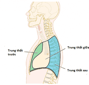
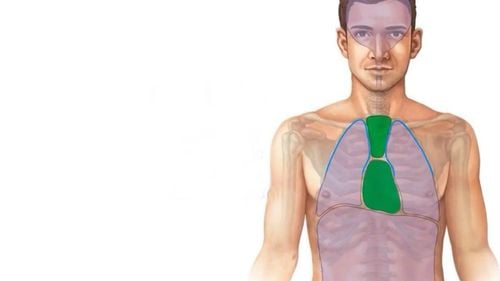
The mediastinum is a space located in the center of the thorax, between the pleural spaces. The mediastinum contains important organs such as the heart, great vessels, trachea, esophagus, thoracic duct, thymus, cardiac nerve, phrenic nerve, lymph nodes,.... Mediastinal tumor is the most common disease in the group of mediastinal diseases. Mediastinal tumors are common according to certain locations such as:Common tumors in the anterior mediastinum: thymoma, lymphoma, thyroid goiter, parathyroid adenoma,... Tumor common in the mediastinum: tumor trachea, lymphoma Common tumors in the posterior mediastinum: esophageal tumor; tumors, nerve cocoon; Diaphragmatic hernia ; Pancreatic pseudocyst,... Imaging methods such as straight-angle chest x-ray, chest computed tomography with contrast injection, nuclear magnetic resonance imaging (MRI,... help diagnose mediastinal tumors. However, conventional imaging methods cannot determine whether a tumor is benign or malignant. To find out the tumor nature to choose the right treatment method, biopsy is an extremely important method.
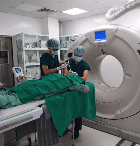
Mediastinal biopsy under computed tomography is a commonly performed method today. Computed tomography helps to accurately determine the location, size, density of the tumor, the anatomical relationship of the tumor to the surrounding organs,... Under the guidance of computed tomography, the technique Biopsy, aspiration of patient samples will be made easier and more accurate. Mediastinal biopsy under computed tomography is a minimally invasive, safe, and highly accurate method. In particular, transthoracic biopsies have an advantage in peripheral thoracic lesions that cannot be reached by endoscopy.

Patient has severe coagulopathy that cannot be controlled. The lesions are located deep in the mediastinum, surrounded by many large blood vessels, the patient has severe lung lesions such as severe alveolar dilatation, lying close to the lungs. Large air cysts can cause pneumothorax.
2. Preparation before mediastinal biopsy under computed tomography
2.1. Executor
The implementation team includes a specialist doctor who is proficient in transthoracic biopsy technique under the guidance of computed tomography, an assistant doctor, a nurse trained in this technique, and an electro-optical technician. If the patient cannot cooperate, more doctors and anesthesiologists are needed.2.2. Means required
Computer tomography machine; film, film printers and image storage systems. Anesthesia, anesthetic (if indicated), antiseptic, water-soluble iodinated contrast agent, medicine and first aid kit, specialized biopsy needle, syringe, cotton, bandage, gauze .2.3. Prepare the patient
To prepare for a transthoracic biopsy, the patient and family will be explained by the medical staff the purpose and possible complications when conducting the biopsy. Biopsy is performed only if the patient consents and signs the consent form for the procedure.The doctor will explain the steps for the patient to cooperate. Especially when it is necessary to lie still, exhale fully and hold the breath of the needle as well as when withdrawing the biopsy needle. When the patient breathes, the mediastinal tumor will also be moved with breathing. Therefore, maintaining the same breath hold each time will help the biopsy needle to be inserted into the correct lesion site.
Patients need to inform the doctor about the drugs they are taking. The doctor may ask the patient to stop some medications a few days before the transthoracic biopsy, especially anticoagulants, antiplatelet drugs, etc. The patient also needs to inform the doctor about the other diseases and conditions, if any.
The doctor appoints to perform subclinical tests and re-examine the hemodynamic, respiratory, heart rate,... to ensure that the patient has a suitable health condition to conduct a routine biopsy. chest wall.
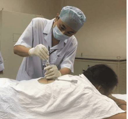
3. Steps to conduct mediastinal biopsy under computed tomography
Local anesthetic is performed with 2% Lidocaine, the amount of anesthetic is from 2-10ml depending on the biopsy site. The patient is brought to the computerized tomography table, placed in a suitable position, exposing the entire chest. Depending on the location of the injury, the patient will be placed in a supine, prone, or prone position. However, the supine or prone position is the most commonly used position, because in these positions the patient is usually still, with little movement. Besides, the biopsy lesion is located in the mediastinum, the puncture directions are mainly from anterior and posterior to the mediastinum, but rarely from the sides. During the entire procedure, from the positioning scan to the biopsy, the patient must remain completely in one position.Positional imaging to determine the expected location of the biopsy. Move the cut line marker to the upper limit of the intended biopsy site on the patient's chest wall. Stick the local needle needle on the site to be biopsied. Then, the doctor ordered a CT scan of the area where the needle was glued.

Disinfect the biopsy area. The doctor washes his hands, puts on a shirt, wears gloves, and puts a sterile sheet with holes on the required position. Local anesthetic layer by layer, then insert the guide needle according to the intended puncture, CT scan to determine the path and position of the needle.
When the guide needle has entered the right lesion (mediastinal tumor), the doctor withdraws the needle lumen, inserts the cutting tool and proceeds to cut the specimen.
After done, the doctor withdraws the biopsy needle, collects the specimen in the needle, and fixes the specimen for anatomical examination. The nurse bandages the puncture site.
After the procedure, the patient is immediately taken into the computed tomography class to detect early complications of pneumothorax or pleural bleeding. The patient was closely monitored for his general condition, chest pain, shortness of breath, and hemoptysis 24 hours after transthoracic biopsy. After 1 day, the patient will have a straight chest X-ray to check for pneumothorax and bleeding.
4. Complications after mediastinal biopsy under computed tomography and how to handle
Bleeding at the puncture site: Treat by compressing the puncture site.Coughing blood : treatment depends on the level.
If coughing up little blood, have the patient lie down on the bed, breathe oxygen, take cough suppressant. If coughing from 20ml or more, have the patient lie down on the bed, head low, face turned to one side, breathe oxygen. Intubate mechanical ventilation if the patient shows signs of respiratory failure. If there is pneumomediastinum or complications due to punctures of dangerous organs and structures in the mediastinum, the doctor will handle it on a case-by-case basis.
Mediastinal biopsy under computed tomography is a common imaging method today. Computed tomography helps to accurately determine the location, size, density of the tumor, the anatomical relationship between the tumor and the surrounding organs. However, for accurate results, patients need to find a reputable medical facility to perform.

Please dial HOTLINE for more information or register for an appointment HERE. Download MyVinmec app to make appointments faster and to manage your bookings easily.


