This is an automatically translated article.
The article was professionally consulted with Specialist Doctor I Nguyen Dinh Hung - Radiologist - Department of Diagnostic Imaging - Vinmec Hai Phong International General Hospital.And Master, Doctor Nguyen Le Thao Tram - Doctor of Diagnostic Imaging - Department of Diagnostic Imaging - Vinmec Nha Trang International General Hospital.
Imaging diagnosis of mediastinal syndrome on radiographs includes signs describing the presence of abnormal cells or foreign pathology in the mediastinum, including: air, fluid, tumor, or tumor. lime close.
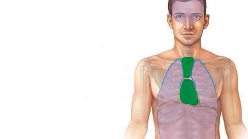
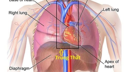
1. Anatomical features of mediastinum
Right mediastinal border includes:
venous trunk - right cephalic arm. Superior and inferior vena cava. Right atrium. Right lung hilum. The left mediastinal border includes:
Left subclavian artery. The aortic arch and the pulmonary arch. Left ventricle. Left lung hilum. Mediastinal compartments:
Anterior mediastinum is limited by the posterior border of the sternum anteriorly, posteriorly by the anterior border of the heart. The mediastinum is limited from the anterior border of the heart to the anterior aspect of the spine, and includes organs such as the esophagus, trachea and main bronchus, superior and inferior vena cava, the X nerve, and the recurrent nerve. reverse larynx. The posterior mediastinum is limited from the anterior aspect of the spine to the posterior aspect of the spine, and includes the following structures: the descending aorta, the single and semi-simple veins, the vertebral bones, and spinal related structures. The superior mediastinum is located at the top, and includes the thyroid gland, esophagus, trachea, aortic arch, and great blood vessels.
2. X-ray features of mediastinal syndrome
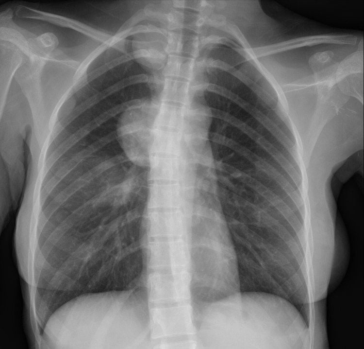
Identify mediastinal lesions on radiographs by the following basic features:Clear, continuous outer border, unclear inner border. Create obtuse angle with mediastinum. There is no bronchogram. Imaging signs in imaging diagnostics of mediastinal lesions:

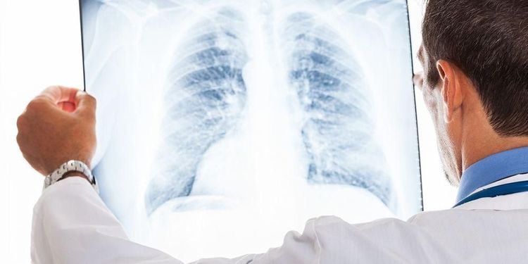
Chụp X-quang giúp chẩn đoán các tổn thương trung thất
2.1. Shadow sign
If the opacity partially or completely obliterates the cardiac margin, the lesion is in the anterior, medial or lingual lobe of the lung, the anterior mediastinum, or the anterior portion of the pleural space. If the opacity overlaps but does not obliterate the cardiac margin, the abnormality is located posteriorly, in the lower lobes of the lung, the posterior mediastinum, or the posterior portion of the pleural space.
2.2. Neck and chest sign
This is a sign that the lesion is located in the superior mediastinum, if the superior border passes beyond the superior border of the clavicle, it is located in the posterior mediastinum, if it does not go beyond the clavicle, the abnormality is in the anterior mediastinum.2.3. Chest and abdomen sign
This finding helps determine if the lesion is located in the chest (mediastinum) or in the abdomen. If the opacity on the radiograph has a clear bottom when it passes through the diaphragm (air nature), the lesion is thoracic (mediastinum), otherwise, it is likely to be in the abdomen.2.4. Signs of covering the hilum
If the lesion covers the hilum, but within the opaque mass, there is still the presence of the pulmonary artery, the lesion is definitely not in the hilum. At that time, it is necessary to further determine whether the cardiac margin is erased, if so, the lesion is located in front of the pulmonary artery, otherwise, behind the pulmonary artery.2.5. Signs of vascular convergence
This sign helps to distinguish whether the opacity is due to an enlarged pulmonary artery or a mediastinal tumor
If the vessel is directed toward the center of the shading, the vessel stops at the border or comes out of the opacity by no more than 1 cm, think to many lesions that are due to large pulmonary arteries. If the vessel points toward a non-central point, think of the lesion as a mediastinal tumor. Accordingly, doctors can diagnose mediastinal tumors based on the location of appearance: goiter, goiter, malformed tumor, lymphoma, hernia through the foramen of Morgagni, vasocardial cyst, bronchial cyst , Transesophageal hiatus hernia, Esophageal tumor, Aortic aneurysm, Bochdalek foramen hernia, Neuroma.
Diagnostic imaging of mediastinal syndrome on radiographs helps doctors detect many diseases, thereby helping patients to have timely examination and treatment. However, patients should also choose reputable examination sites, because this technique requires highly skilled operators and modern machinery to produce the clearest X-rays.

Chụp X quang có thể chẩn đoán bệnh u trung thất
The general hospital uses the new generation of mobile and fixed digital X-ray techniques of the world's leading medical equipment manufacturers such as: GE, Toshiba.... as well as current techniques other tools to diagnose and treat lung diseases.
With modern facilities of international standards, quality medical services, a team of medical professionals, experienced doctors will bring optimal treatment results to customers.
Friendly hospital environment, international standard pre-, post-operative care facilities will bring the most accurate diagnosis results, best treatment, fastest health recovery.


