This is an automatically translated article.
The article was professionally consulted with Specialist Doctor I Le Van Quang - Department of General Surgery - Vinmec Nha Trang International General HospitalMediastinoscopy is an invasive surgical method that allows doctors to perform biopsies to conduct adjunctive diagnostic tests for mediastinal diseases, including mediastinal lymphadenopathy.
1. 1. What is mediastinoscopy?
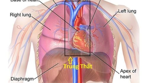
Mediastinoscopy is a minimally invasive surgical method (surgery with minimal trauma and minimal damage to the body) by opening an opening into the chest cavity for visualization. The mediastinum includes the heart, trachea and esophagus, nerves, lymph nodes, and the thymus. These are organs of the immune system that help the body fight infections.
Perform mediastinoscopy by making a very small incision in the chest and creating an entrance to insert the mediastinoscope into the chest. This bronchoscope has a camera attached to transmit images to a screen to help doctors and surgeons assess the condition inside the chest.
When observing the endoscopic image, the surgeon can conduct some more tests if needed. Through the laparoscope, the surgeon can also remove a small sample of the patient's chest and examine it under a microscope to help diagnose the disease.
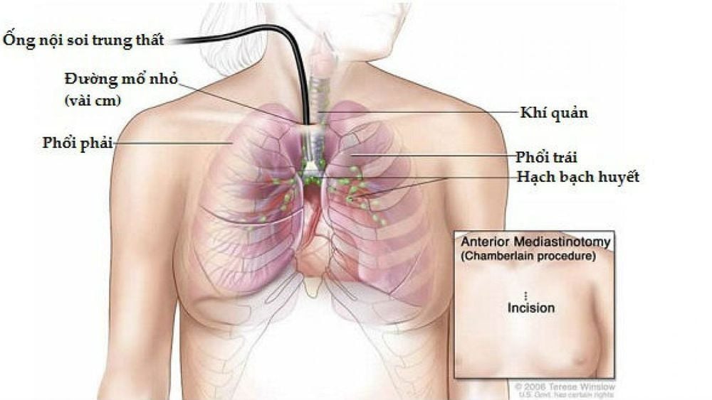
Nội soi trung thất
2. Purpose of mediastinoscopy
Mediastinoscopy is done for the following purposes:
Evaluation of problems of the lungs and mediastinum, eg sarcoidosis; Diagnosis of lung cancer or lymphoma (lymphoma). Mediastinal lymphadenopathy on CT alone or with lung tumor, mediastinal tumor but no histological diagnosis; Evaluation of metastatic status of the lymph node group in the mediastinum before pneumonectomy in the treatment of lung cancer; Helping doctors decide on the optimal treatment and prognosis for lung cancer; Diagnose other infectious diseases involving the lungs, eg tuberculosis .
3. How is mediastinoscopy performed?
Before surgery, the patient is placed infusion line in the hand to administer fluids and drugs; The patient is then anesthetized to sleep during the operation; After the patient is asleep, the doctor performs an intubation - a small tube in the windpipe to control breathing during surgery; Next, the doctor disinfects the surgical area in the neck and chest, makes a small incision (about a few centimeters) in the chest wall, above the breastbone; Through the incision, the doctor or surgeon inserts the mediastinoscope into the chest. The camera attached to the end of the endoscope will transmit images to the screen so that the doctor or surgeon can see the inside of the chest; During endoscopy, if necessary, the surgeon can also insert some other specialized instruments by other routes; The doctor examines the spaces between the heart and lungs, and biopsies the lymph nodes or tissues if there are abnormalities to perform testing and evaluation; After withdrawing the mediastinoscope from the thorax, the doctor stitched and closed the incision. The whole process takes about an hour.
4. Does mediastinoscopy cause complications?
Most cases of mediastinoscopy do not cause complications. Sometimes, the endoscope can cause temporary nerve damage and cause a patient to develop hoarseness, which in some cases is irreversible.
In rare cases, the patient may experience blood loss and require a blood transfusion because of the significant blood loss, and may need to be converted to open surgery. In this case, the patient may have a pneumothorax (ie air leaks out from the lungs) so the patient needs to be intervened by placing a drain between the two ribs for a few days.
In addition, in very rare cases, mediastinoscopy can cause atelectasis or esophageal tear.
If you have one of the following problems, the patient should be re-examined immediately:
Bleeding at the incision site; High fever, hoarseness that lasts for days or gets worse; Severe chest pain, neck swelling; Difficulty breathing, difficulty swallowing.
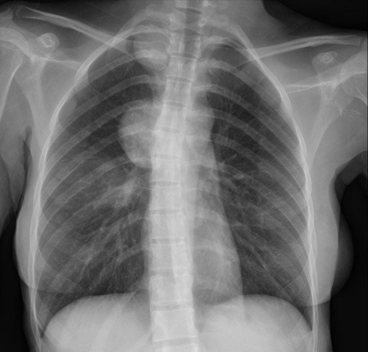
Hình ảnh hạch trung thất trên CT
5. How to prepare and monitor before and after mediastinoscopy?
5.1 Before mediastinoscopy The doctor examines and evaluates and discusses before surgery; If the patient is taking anticoagulants, they will be asked to stop taking them a few days in advance; Patients are also asked to fast and fast for a period of time before surgery. 5.2 After mediastinoscopy The patient can go home after regaining consciousness and drinking water without choking or choking. However, some cases will require a stay in the hospital for a few days; If surgery by sutures does not dissolve, the patient needs to be re-examined to remove the sutures; You may have a mild sore throat and pain at the incision site for a few days after surgery. Mediastinoscopy is a non-invasive surgical technique indicated for the evaluation and diagnosis of lung and mediastinal pathologies, has the ability to perform widely and safely, and has diagnostic value with high accuracy. .
Please dial HOTLINE for more information or register for an appointment HERE. Download MyVinmec app to make appointments faster and to manage your bookings easily.


