This is an automatically translated article.
Post by Master, Doctor Mai Vien Phuong - Department of Examination & Internal Medicine - Vinmec Central Park International General Hospital
Esophageal cancer (EC) is a common malignancy of the gastrointestinal tract and originates in the epithelium of the lining of the esophagus. It has been confirmed that early esophageal cancer lesions can be cured by endoscopic therapy and that the therapeutic effect is comparable to surgery. Upper gastrointestinal endoscopy remains the gold standard for diagnosing esophageal cancer.

1. Real-time diagnosis of esophageal squamous cell carcinoma
Guo et al have developed a new computer-aided diagnostic system system based on SegNet architecture that can perform real-time automated diagnosis of precancerous and cancerous lesions early esophageal squamous cell epithelium in conditions other than magnifying and magnifying endoscopy. Data from four medical centers in three countries including China, the United States and India were collected and used for training a new computer-aided diagnostic system model. Four data sets from primary and noncancerous esophageal squamous cell carcinoma including still images and real-time video were used to verify their effects. The system's SEN and SPE are 98.04% and 95.03% for still images, respectively. In 27 unmagnified videos, the SEN per frame and per lesion were 60.8% and 100%, respectively. In 20 magnified videos, the SEN per frame and per lesion were 96.1% and 100%, respectively. The unchanged full-range normal esophageal videos included 33 videos (SPE 99.9% per frame, SPE 90.9% per case).
In another multicenter, case-control, diagnostic study, Luo et al developed and validated the Gastrointestinal Artificial Intelligence Diagnostic System (GRAI DS) for the diagnosis of gastrointestinal cancer upper gastrointestinal tract through analysis of clinical endoscopy imaging data. Six hospitals of different levels of experience in endoscopic diagnosis of upper gastrointestinal cancer in China participated in the study. More detailed data on early esophageal cancer are to be expected. The latest research results on early esophageal cancer detection in real time are from Fukuda et al. They used 23746 images of 1544 cases of pathologically confirmed superficial esophageal squamous cell carcinoma and 4587 images of 458 noncancerous cases and normal tissues to build the model. CNN-SSD deep.

2. Accuracy of artificial intelligence in real-time diagnosis of esophageal squamous cell carcinoma
NBI or BLI technology was used to collect 5-9 second video clips of 144 patients as verification dataset. SEN, SPE, and accuracy for artificial intelligence were 86%, 89%, and 88%, respectively, and 74%, 76%, and 75% for experts, respectively. This is an important study that paves the way for the development of better models to detect esophageal cancer lesions in a real-time clinical setting, but randomized prospective clinical trials are needed. Verification. Another research team from Japan has developed an artificial intelligence system to calculate the invasive depth of esophageal squamous cell carcinoma. In total, 23977 images, including WLI and NBI/BLI images of pathologically proven esophageal squamous cell carcinoma from endoscopic video and still images, were collected as a single volume. learning data. An independent validation dataset of 102 video images of esophageal squamous cell carcinoma was performed in the study. Two types of video, non-magnified endoscopy with WLI and magnified endoscopy with NBI/BLI from 4-12 seconds, were included. The CNN model was compared with 14 experts in the field of endoscopy. Accuracy, SEN and SPE performance for AI outperforms expert performance in both video types. The artificial intelligence model was concluded to be effective in real-time measurement of the depth of invasion of esophageal squamous cell carcinoma.3. Artificial intelligence in detecting Barrett's esophagus and esophageal adenocarcinoma
Barrett's esophagus is a precursor to adenocarcinoma of the esophagus. The prevalence of adenocarcinoma of the esophagus is increasing rapidly and the prognosis of advanced cases is poor. However, early detection of cases that can be treated promptly by endoscopic methods has a chance of cure. Because it is difficult to detect early endoscopic tumors, early lesions are often missed. This is because Barrett tumors are usually initially flat, with only minor changes in the color and texture of the mucosa. These changes can be seen in HD-WLI made by experts. However, because of the low rate of advanced tumor formation (<1%/patient year), many general endoscopic surgeons rarely encounter early-forming Barrett tumors, thus they are unfamiliar with endoscopic morphology. of it and the lesions are not recognizable. To improve screening for esophageal adenocarcinoma and increase SEN and speed of examination, artificial intelligence-assisted endoscopy is of great value. In addition, it can also provide convenience for the endoscopist facing challenges and concerns, so as not to miss early lesions.

Diagnosis of esophageal adenocarcinoma
In 2013, van der Sommen et al. first proposed an SVM algorithm for early cancer detection in esophageal adenocarcinoma using imaging HD endoscopy, color histograms, and simple statistical data on Gabor features, with a classification accuracy of 95.9% and an AUC of 0.992. They then found that the system detected 36 from 38 lesions with a recall of 0.95 and an accuracy of 0.75 through clinical validation. In 2016, van der Sommen and colleagues tested a computer algorithm (automatic image recognition system) that uses specific textures, color filters, and machine learning to detect cancerous lesions. Early on Barrett's esophagus based on specific imaging details. All images are of high perceived quality. The algorithm was developed and tested using 100 endoscopic images of 44 patients with Barrett's esophagus. Primary tumor lesions were identified in per-image analysis, with SEN and SPE of 0.83. At the patient level, the SEN and SPE of the system were 0.86 and 0.87, respectively. Swager et al performed the first study based on direct histologically correlated volumetric laser endoscopic (VLE) imaging in 2017, which developed a clinically inspired computer algorithm for Barrett's esophagus. VLE is an advanced imaging system that can scan the esophageal wall up to 3 mm deep with near-microscopic resolution. Total 60 VLE images from high quality ex vivo VLE histological correlation database used in the study, including 30 nondysplastic Barrett esophagus and 30 HGD/esophageal adenocarcinoma images Soon.
Algorithm of feature "classification statistics and signal decay"
Algorithm of feature "classification statistics and signal decay" shows good performance in detecting Barrett's esophagus tumor (AUC = 0.95), and thus it can assist endoscopists in detecting early tumors on the VLE. Van der Sommen et al also compared the performance of the new computer-aided diagnostic system methods with the two VLE experts' accuracy classification, with a maximum AUC in the range of 0.90. -0.93 for the machine learning approach compared to an AUC of 0.81 for medical professionals. Horie et al demonstrated that CNN's diagnostic accuracy for esophageal adenocarcinoma was 90% (19/21). Four lesions of esophageal adenocarcinoma were missed, possibly due to inadequate educational imaging for esophageal adenocarcinoma. Only eight were used in the study.
Diagnosis of depth of invasion of esophageal adenocarcinoma
Some studies question whether surgery should always be used to treat esophageal adenocarcinoma with deep submucosal invasion are not. However, it is accepted that depth of invasion is at least one of the greatest risk factors for metastasis. In the search related to the term “depth of infiltration/infiltration of esophageal adenocarcinoma”, no published articles were found. Fortunately, research on artificial intelligence penetration depth measurement for gastric and colon cancer has been published, which may be worth learning for research on cancer penetration depth. esophageal glandular epithelium. In the study by Zhu et al., a total of 790 WLI were used as the development dataset and another 203 images as the experimental dataset and the new computer-aided diagnostic system-CNN system, was developed from ResNet50, used to determine the depth of invasion of gastric cancer. At the cutoff value of 0.5, the SEN was 76.47%, the SPE was 95.56%, and the overall accuracy was 89.16%. The artificial intelligence system achieves significantly higher accuracy than human endoscopes.
New AI-based computer-aided diagnostic system designated to improve the quality of Barrett's esophagus
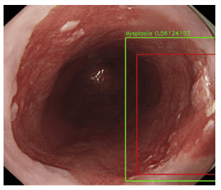
New AI-based computer-aided diagnostic system is indicated to improve the quality of the assessment of Barrett's esophagus, especially for non-expert endoscopists. In 2019, Trindade and colleagues reported an artificial intelligence software called intelligent real-time image segmentation for endoscopic surveillance of Barrett's esophagus. Intelligent real-time image segmentation identified three previously established VLE features associated with histological dysplasia and visualized them using different palettes superimposed on the image VLE. A multicenter randomized controlled trial (NCT03814824) is underway to further validate the artificial intelligence system. In another study, de Groof et al evaluated the preliminary diagnostic accuracy of a new computer-aided diagnostic system in detecting Barrett's cancer during rectal endoscopy. next. The laboratory dataset included direct endoscopic procedures of 10 patients with nonspasmodic Barrett's esophagus and 10 patients with confirmed Barrett's cancer. WLI at each 2 cm level of the Barrett segment was collected and analyzed. The accuracy, SEN and SPE of the new computer-aided diagnostic system are 90%, 91% and 89%, respectively. These results suggest that the new computer-aided diagnostic system is ready to be tested in larger multicenter trials.
Please dial HOTLINE for more information or register for an appointment HERE. Download MyVinmec app to make appointments faster and to manage your bookings easily.
References
Liu Y. Artificial intelligence-assisted endoscopic detection of esophageal neoplasia in early stage: The next step? World J Gastroenterol 2021; 27(14): 1392-1405 [DOI: 10.3748/wjg.v27.i14.1392]. Merkow RP, Bilimoria KY, Keswani RN, Chung J, Sherman KL, Knab LM, Posner MC, Bentrem DJ. Treatment trends, risk of lymph node metastasis and outcome of localized esophageal cancer. J Natl Cancer Inst . 2014; 106 . [ PubMed ] [ DOI ] Liu Y. Artificial intelligence-assisted endoscopic detection of esophageal neoplasia in early stage: The next step? World J Gastroenterol 2021; 27(14): 1392-1405 [DOI: 10.3748/wjg.v27.i14.1392]


