This is an automatically translated article.
The article is professionally consulted by MSc, BS. Dang Manh Cuong - Doctor of Radiology - Department of Diagnostic Imaging - Vinmec Central Park International General Hospital.Ultrasound is a safe and commonly used diagnostic imaging technique. In which, Duplex ultrasound is a combination of normal and Doppler ultrasound, very effective in diagnosing cardiovascular diseases in general, as well as venous disease in particular.
1. What is Duplex Ultrasound?
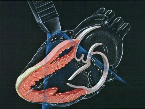
Duplex ultrasound is considered a special technique that combines basic ultrasound with Doppler ultrasound. This ultrasound technique allows to examine the morphology and structure of blood vessels, as well as to evaluate the movement of blood flow in the body.In which, cardiovascular diseases are diseases related to blood vessels such as veins, arteries and capillaries. Therefore, Duplex ultrasound is very effective in diagnosing cardiovascular diseases in general, as well as venous diseases in particular.
2. What is Duplex ultrasound used for?
Duplex Ultrasound is commonly used to evaluate blood flow in various arteries and veins.2.1 Varicose veins of the lower extremities Ultrasound technique will help to evaluate the reverse flow in the superficial veins, the deep veins located between the muscle layers of the legs and even the perforating and intervening veins shallow and deep.
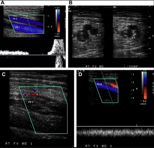
Siêu âm Dopplermàu cắt dọc Tĩnh mạch đùi có hai nhánh (FV)
Besides, ultrasound is also capable of measuring the size of superficial varicose veins, doctors will mark on the surface of the skin corresponding to the location of the varicose veins, through which the doctor will proceed Vascular surgery can remove these pathological veins with various interventional methods.
2.2 Deep Vein Thrombosis Deep vein thrombosis is a condition in which a blood clot forms in the lumen of a vein. These blood clots over time will grow larger, causing venous flow obstruction, causing leg edema and can float to the lungs causing pulmonary embolism. After that, the blood clots will gradually shrink, fibrosis and stick to the vein wall, making the vein wall thicken, the valves can be damaged, lose function... post-thrombotic insufficiency. In the acute phase, Duplex ultrasound will help diagnose varicose veins, blood clots in the venous lumen, or provide images of veins that cannot be deflated as usual. In the chronic stage, Duplex ultrasound will show thicker venous walls, much fibrosis, venous occlusion, incompletely collapsed veins, and prolonged retrograde flow in the deep veins. In addition, Duplex ultrasound is also a technique to help doctors diagnose a number of other less common venous diseases such as aneurysms, varicose veins, venous compression, venous wall tumors...

Hình ảnh kết quả siêu âm tĩnh mạch gan
3. How does the Duplex ultrasound procedure work?
You lie in bed and the sonographer places an ultrasound transducer on your skin at the part of the body that needs to be examined. Lubricant is applied to your skin to help the transducer make good contact with your body. The transducer is connected directly to the machine by a wire through which information is quickly transferred to the screen. The transducer emits ultrasound waves that, through the skin, enter your body. The ultrasound waves that are then reflected back from various structures in the body are picked up by the transducer, transmitted through the wire, and into the ultrasound machine. They are seen as images on the screen. This image is constantly updated so Duplex ultrasound can show movement of structures. For example, the heart valves open and close during echocardiography. The sonographer can move the transducer across the surface of the skin for different viewing angles. The ultrasound procedure is painless and can take about 15-45 minutes, depending on the parts that need to be examined. The resulting ultrasound image can be saved as a single image or by video.
Các sóng siêu âm đi xuyên qua da và phản hồi lại các tín hiệu truyền tải về máy siêu âm
4. Does the ultrasound test have any side effects or complications?
Ultrasound is completely safe and painless. Unlike X-rays and other imaging techniques, ultrasound does not use radiation. No problems or complications due to ultrasound have been found so far.Duplex ultrasound is a reliable, safe, easy to perform imaging test in the diagnosis and treatment of venous diseases. However, this is a diagnostic technique whose results depend largely on the skill and experience of the operator. A well-trained, experienced sonographer will give you a more reliable ultrasound result.
Before taking a job at Vinmec Central Park International Hospital from December 2017, Doctor Dang Manh Cuong has over 18 years of experience in the field of ultrasound - diagnostic imaging in Transport Hospitals. Hai Phong, MRI Department of Nguyen Tri Phuong Hospital and Diagnostic Imaging Department of Becamex International Hospital.
Any questions that need to be answered by a specialist doctor as well as customers wishing to be examined and treated at Vinmec International General Hospital, you can contact Vinmec Health System nationwide or register online HERE.