This is an automatically translated article.
The article is professionally consulted by BSCK II Nguyen Quoc Viet - Department of Medical Examination & Internal Medicine - Vinmec Danang International General Hospital.
Echocardiography is one of the most common, non-invasive diagnostic methods used to evaluate cardiac morphology, function, and etiology of structural heart disease.
1. What is echocardiography?
Echocardiography is a simple, reliable, non-invasive method to investigate the morphology, function and hemodynamics of the heart chambers, septum, pericardium, and major vessels connected to the heart. heart.
Echocardiography method will help doctors in diagnosing heart health quickly and effectively. One of the fastest, safe, and non-invasive diagnostic methods to provide timely treatment to help maintain a healthy heart.
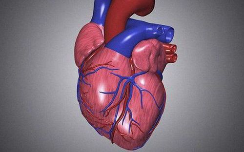
The doctor uses an echocardiogram (harmless to health) transducer to observe the structure of the heart and check how the heart is working. What it means for an echocardiogram to help your doctor find out:
The size and shape of your heart; the size, thickness, and movement of the heart's walls How the heart moves How the heart's pumping power Is the heart valves working properly Is blood leaking back through the heart valves (reflux) Is the heart valve damaged narrow or not There is no tumor or inflammatory mass around the heart valve. In addition, an echocardiogram helps doctors find:
Problems with the pericardium Problems with the great blood vessels going and leaving the heart Blood clots in the chambers of the heart Abnormal holes between heart chambers.
2. Abbreviated symbols in echocardiogram results

Siêu âm tim tại Bệnh viện đa khoa Quốc tế Vinmec
Ao : Aorta LA : Left Atrium RA : Right Atrium LV : Left Ventricular RV : Right Ventricular LVOT : Left ventricular ejection chamber outflow tract) RVOT: right ventricular outflow tract EF: ejection fraction EF (teich) ejection fraction according to Teicholz method IVSd: Interventricular Septal thickness Diastolic) IVSs : Interventricular Septal Systolic Thickness LVEDd : Left Ventricular End Diastolic Dimension LVEDs : Left Ventricular End Systolic Dimension LVPWd: Degree left ventricular posterior wall diastolic (Left ventricular posterior wall diastolic) LVPWs: left ventricular posterior wall systolic (Left ventricular posterior wall systolic) EDV (Teich): End diastolic volume according to Teicholz method (End diastolic Volume) ESV (Teich): End-systolic volume in the Method Teicholz (End-systolic volume) SV (Teich) : Stroke Volume Ann : Anterior mitral valve leaflet AML : Anterior mitral valve leaflet PML : Posterior mitral valve leaflet ) MVA : Mitral valve area diameter PHT : Pressure half time TV : Tricuspid Valve AnnTV : Annular Tricuspid Valve diameter AV : Aortic Valve AoVA: Diameter of aortic annulus AoR : Diameter of Valsalva sinus STJ : Sinus-tubal junction AoA : Ascending aorta AoT : Aortic arch AoD : Descending aorta AVA : Diameter of the aortic valve hole.
Echocardiography is one of the important methods to ensure a healthy heart, but patients need to choose methods that require high technology.
Vinmec International General Hospital is one of the hospitals that not only ensures professional quality with a team of leading medical professionals, modern equipment and technology, but also stands out for its examination and consultation services. comprehensive and professional medical consultation and treatment; civilized, polite, safe and sterile medical examination and treatment space.
Please dial HOTLINE for more information or register for an appointment HERE. Download MyVinmec app to make appointments faster and to manage your bookings easily.