This is an automatically translated article.
The article was professionally consulted with Specialist Doctor I Nguyen Thi Bich Nhi - Ophthalmologist - General Surgery Department - Vinmec Nha Trang International General Hospital.The evoked potential is the potential recorded after stimulating the central nervous system with organ-specific stimulation waves. Visual evoked potentials use light stimuli to assess the integrity of optic nerve transmission, from the retina to the occipital visual cortex.
1. Concepts and things to know about evoked potentials
1.1 What is a evoked potential? An evoked potential (Eps) is an electrical brain wave recorded after a stimulus specific to the responses recorded from the CNS or a wave produced by CNS stimulation.In the past, evoked potentials were very important, but due to the appearance of MRI, now the role of this method is much narrowed. However, there is still a huge difference between MRI and evoked potentials.
Brain MRI provides information about the anatomical structure of the nervous system, and evoked potentials provide information about the function of neurotransmitters in the brain.
Disadvantages of MRI are high cost, difficult to perform many times and cannot be done at the hospital bed. Meanwhile, evoked potentials are cheaper, can be performed at the bedside and can be repeated many times.
Stimulation-evoked potential recording allows recording and analysis of electrical waves emitted by the cerebral cortex and spinal cord. Stimulus potential waves will occur if there is a central nervous system response to electrical stimulation of the peripheral nerves or when stimulating the sensory organs (visual, auditory). Except for motor excitation potentials, recording evoked potentials usually requires several hundred to several thousand times of stimulation, using a computerized recorder to store the received signals. The signals will be averaged and noise removed, so that the excitation potential waveforms will be clearly recorded. The evoked potentiometers are accompanied by specialized stimuli such as light, sound or magnetic fields.
1.2 Types of evoked potentials Commonly used types of evoked potentials include:
Somatosensory evoked potentials (SEPs): Obtained by stimulating afferent nerve fibers of the muscles and skin. Somatosensory potentials help examine the peripheral nervous system, especially the proximal extremities; detect central sensory nerve damage; evaluate the damage caused by surgery such as dissection of the inner layer of the carotid artery so that it can be repaired in time. Brainstem auditory evoked potentials (BAEPs): Use sound to generate stimulus waves to assess the function of nerves and auditory pathways in the brain stem. Visual evoked potentials (VEPs): Use light to generate stimulation waves to assess the function of nerves and visual pathways in the brain stem. Motor evoked potentials (MEPs): Recorded in muscles, peripheral nerves, or spinal cord during cortical or pyramidal stimulation, helps in the diagnosis of motor neuron disease, pyramidal injury, and prognosis in patients with stroke. stroke. Intellectual evoked potential (P300): Measures the latency and amplitude of P300 - reflects the brain's intellectual function. Psychogenic potentials are commonly used in the study of Alzheimer's disease and other dementias, depression, schizophrenia, obsessive-compulsive disorder, attention deficit disorder, and epilepsy in children.

Ghi điện thế gợi kích thích cho phép ghi lại và phân tích các sóng điện phát ra từ vỏ não và tủy sống
2. Concepts and things to know about visual evoked potentials
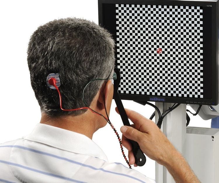
2.1 What is a visual cue potential? Visual evoked potentials (VEPs) are electrical waves of the brain recorded by light stimulation entering the eye that reflect the function of the optic nerve and the integrity of the visual pathway, from the retina to the eye. occipital visual cortex.2.2 How to perform the visual evoked potential The patient is in a relaxed position. Stimulating light into one eye. The light is in the form of a checkerboard, consisting of black and white squares that are maximally contrasting and alternating. Light has a frequency of about 2 Hz.
The evoked potential recording electrode consists of an electrode in the center of the occipital and 2 electrodes on the sides 5cm apart. Do about 100 to 200 times of stimulation. There are usually two waves, N75 (negative wave with a latent time of 75ms) and P100 (the next positive wave has a latent time of 100ms). The most important visual cue potential is P100 because it reflects the integrity of the visual pathway.
2.3 Application of optic potentials Visual evoked potentials help assess the integrity of the optic nerve pathway from the second cranial nerve, through the optic tract and optic tract, to the lateral iliac and projection bodies through the striatum to the visual cortex. Visual evoked potentials are more sensitive than MRI in examining pre-interfocal lesions, but can also help differentiate post-interference visual pathways.
Visual evoked potentials may show abnormalities in multiple sclerosis and cortical blindness, glaucoma (hyperhydramnios), Parkinson's disease. If clinical and MRI findings show spinal cord injury and visual evoked potentials are abnormal (although MRI does not reveal second cranial nerve abnormalities and the patient does not complain of amblyopia) then it should be suspected. multiple sclerosis or Devi's disease.
In addition, visual evoked potentials are used to measure visual acuity in young children.
Please dial HOTLINE for more information or register for an appointment HERE. Download MyVinmec app to make appointments faster and to manage your bookings easily.


