This is an automatically translated article.
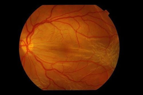
If left untreated, high blood pressure can lead to complications in many other organs such as the heart, brain, kidneys, blood vessels, etc. Similar to diabetes, high blood pressure can cause damage to small blood vessels. in the retina. High blood pressure can cause blockage of blood vessels in the eye, bleeding in the eye, papilledema... affecting the patient's vision.Vascular manifestations in the retina indirectly reflect vascular changes and damage in organs such as the heart, kidney, and brain because the vascular structure of these organs is similar. In fact, clinicians can base on retinal changes to assess the extent and response to treatment of hypertension.
In the early stages, retinopathy does not affect vision much, so it is easy to ignore. In the later stages, when there are serious injuries and complications on the retina, the eyes will be blurred, difficult to treat and difficult to recover.
1. Retinopathy due to hypertension
1.1 Arterial narrowing This is a manifestation of increased vascular pressure. In severe hypertension, precapillary arterioles are blocked, forming a soft, cottony discharge.
Xuất tiết mềm dạng bông
Xuất huyết hình ngọn nến
Sao hoàng điểm
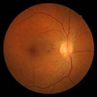
Xơ cứng mạch máu (động mạch và tiểu động mạch)
2. Choroidal disease
Choroidal occlusion: manifested by Elschnig nodules.
Nốt Elschnig
Vệt Siegrist
Bong thanh dịch
2. Optic neuropathy
Optic disc edema, periapical hemorrhage.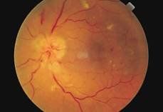
Bệnh lý thị thần kinh
4. Stages of hypertensive retinopathy
Stage I : Arterioles miniaturization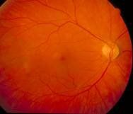
Tiểu động mạch thu nhỏ
cross section
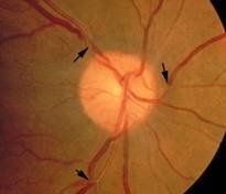
Tiểu động mạch thu nhỏ, lệch hướng tĩnh mạch ở đoạn bắt chéo
Small arterioles like copper wire. Salus Gunn's sign: TM shrinks the tip of the tip, deviating at right angles to the cross section of the artery. Accompanied by candle-shaped hemorrhage, soft discharge, and hard discharge.
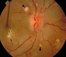
Giai đoạn 3 của bệnh võng mạc tăng huyết áp
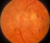
Động mạch thu nhỏ như sợi dây bạc và phù gai thị


