This is an automatically translated article.
The article was professionally consulted by Specialist Doctor I Nguyen Thanh Hai - Doctor of Radiology - Department of Diagnostic Imaging and Nuclear Medicine - Vinmec Times City International Hospital.Cardiovascular magnetic resonance imaging (MRI) can be considered as the most modern, non-invasive, non-X-ray method of cardiac imaging. monitoring cardiovascular diseases in our country.
1. Anatomy of the human heart
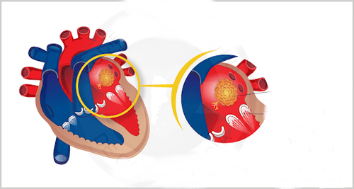
The heart has 4 chambers: 2 upper atria and 2 lower ventricles. The interventricular septum and the atrial septum divide the heart along the longitudinal axis. The heart is located in the mediastinum, above the diaphragm. The outside of the myocardium is surrounded by the pericardium and the inside is lined by the endocardium. The heart is fed by the coronary arteries, which originate from the aorta.The atria of the heart have thinner walls than the ventricles, located at the base of the heart. In particular, the right atrium has an inflow of superior and inferior vena cava, and the left atrium has 4 pulmonary veins.
The ventricles, especially the left ventricle, have a thicker layer of muscle than the atria. The left ventricle is separated from the left atrium by the mitral valve and is responsible for ejecting blood into the aorta to feed the body. The right ventricle and the right atrium are separated by the tricuspid valve, the right ventricle ejects blood into the pulmonary artery for gas exchange in the lungs, then the blood flows through the pulmonary veins into the left atrium.
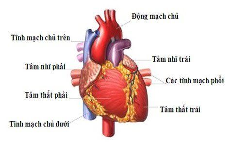
Tim gồm 4 buồng, trong đó 2 tâm nhĩ trên và 2 tâm thất ở dưới
2. What is a cardiac MRI?
Cardiovascular magnetic resonance imaging is a technique that uses a magnetic field and radio waves. The principle of this technique is based on the impact of the waves produced by the MRI machine causing the tissues in the body to absorb and release energy. The cardiac MRI machine then captures the energy, processes it, and converts it into image signals. The image from the MRI machine has the ability to reproduce 3D space, so it is very supportive for disease diagnosis and effective treatment.Cardiovascular magnetic resonance imaging has advantages compared to other techniques such as: high image resolution, high soft tissue contrast, many different cross-sections, no radiation and non-invasive. Therefore, this cardiovascular imaging technique is increasingly widely applied.
3. In what cases is cardiac MRI indicated?
Currently, cardiovascular magnetic resonance imaging is indicated in the following cases:Congenital heart disease to evaluate abnormalities of the flow, anatomy... Cardiomyopathy such as: Myocarditis, dilated myocardium or myocardial hypertrophy, iron deposition in the myocardium, rafted myocardium... Coronary artery disease: Assess the function and mobility of the left ventricle, especially evaluate the survival of the myocardium in infarction acute and chronic blood for which other survey methods are limited; Investigation of infarct and fibrosis areas of the myocardium. Diseases of the pericardium, blood vessels; Heart valve disease ; Primary or secondary cardiac tumor: MRI provides images of tumor nature, so that doctors diagnose, identify and evaluate the extent of tumor invasion; Spectroscopy - Spectroscopy.

Chụp cộng hưởng từ tim mạch giúp phát hiện nhiều bệnh lý ở tim
4. Advantages of MRI compared to other techniques
Cardiovascular MRI does not cause the side effects of CT scans or X-rays, and still allows for abnormalities to be detected - something that other imaging methods cannot do. Give images much faster and more accurate than X-rays in the diagnosis of cardiovascular diseases; Non-invasive and harmful like X-rays.5. Notes on Cardiovascular Magnetic Resonance Imaging
Due to the ability to provide clear and accurate images, many people now abuse cardiovascular magnetic resonance imaging, asking their doctor to let them perform this method.This is a very wrong thinking. Cardiovascular MRI should only be taken when it has been carefully examined by a doctor, has ultrasound and some other imaging tests, but finds it necessary, so the doctor appoints additional imaging. MRI should not be used to screen for cancer because it is unnecessary and quite expensive. On the other hand, MRI is also a diagnostic method by machines, not able to examine the doctor instead. The results obtained after the scan require consultation between the medical examiner and the radiologist to obtain accurate results.
6. Note to the patient when taking an MRI
If the body has metal devices implanted, it is necessary to inform the doctor in advance because the magnetic field of the scanner can damage these devices; Should be taken when prescribed by a cardiologist; The syndrome of claustrophobia, closed, should not take MRI; Do not bring phones, tools or metal medical equipment into the examination room; Contrast in MRI is not toxic to the body but can cause mild allergies with the following symptoms: rash, numbness in limbs, nausea, headache... Dr. Hai has more than 20 years of experience. years of experience in the field of diagnostic imaging, especially in the field of multi-slice computed tomography, magnetic resonance. Currently, the doctor is working at the Department of Diagnostic Imaging and Nuclear Medicine - Vinmec Times City International General Hospital.For medical examination and treatment at Vinmec, please go directly to Vinmec medical system nationwide or register online HERE.


