This is an automatically translated article.
The article was professionally consulted by BSCK II Nguyen Quoc Viet - Interventional Cardiologist - Department of Medical Examination & Internal Medicine - Vinmec Danang International General Hospital.Electrocardiogram is a method to monitor the electrical activity of the heart very popular today, almost all medical facilities use electrocardiogram as a main diagnostic tool for cardiovascular diseases. So what is an electrocardiogram test for, what disease will be detected by reading the electrocardiogram?
1. What is an electrocardiogram?
An electrocardiogram, abbreviated as ECG, is a method of monitoring the activity, speed, and rhythm of the heart. When the heart is working, the heart contracts, it will emit variations of the electric current, at this time the electrocardiogram is a curve that records those variations. Through reading the electrocardiogram, we can know the heart's ability to eject blood, know the rhythm and speed of the heart.Trắc nghiệm: Huyết áp của bạn có đang thực sự tốt?
Huyết áp cao hay thấp đều ảnh hưởng đến tình trạng sức khỏe con người. Để biết tình trạng huyết áp của bạn có thực sự tốt không, hãy làm bài trắc nghiệm sau đây để đánh giá.2. Mechanism of action of electrocardiogram
The human heart has 4 chambers to store and pump blood, the upper 2 parts are called the atria, and the 2 larger lower parts are called the ventricles.Blood follows the veins from the organs in the body to the right atrium, blood from the lungs returns to the left atrium. Squeezing the left atrium pumps blood into the left ventricle, and the right atrium pumps blood into the right ventricle. The right ventricle then squeezes to pump blood through the arteries to the lungs, and the left ventricle squeezes to pump blood down to the body. The heart is able to function at a regular and orderly pace thanks to a system of special conductive cells located in the heart muscle.
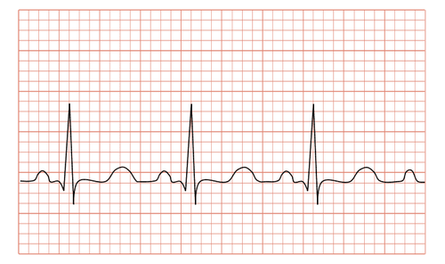
In the right atrium there is a sinus node composed of cells capable of emitting electrical impulses. This electrical impulse is transmitted to the surrounding muscles, causing the two atria to contract (creating a P wave on the electrocardiogram). After that, the current continues to pass along a special cell chain to the atrioventricular node located near the interventricular septum, and then along the Purkinje fiber cell chain running along the interventricular septum and spreading into the surrounding muscles (generating a series of QRS waves). the two ventricles contract. Then, the electrical impulses decrease, the ventricles relax (creating a T wave).
To summarize, the mechanism of action of the electrocardiogram is as follows: cells in the chambers of the heart produce an electrical impulse when the heart is working, these electrical impulses travel through the heart according to a conduction system and are recorded by the electrocardiogram. converted into electrical signals. Some diseases such as arrhythmia, angina can be detected after conducting an electrocardiogram. Therefore, electrocardiogram plays a very important role and is regularly conducted in the hospital.
Cơ chế hoạt động của điện tâm đồ như thế nào?
3. What is an electrocardiogram test for?
An electrocardiogram test records changes in electrical conduction in the heart, thereby helping doctors diagnose conditions such as arrhythmia and conduction disturbances in the heart, myocardial infarction, and anemia. myocardium, heart failure, pericardial effusion, acute pericarditis, chronic heart failure, disturbances of electrolytes in the blood, thickening of the myocardium wall.....Specific readings of the electrocardiogram are as follows:
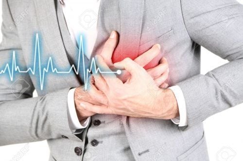
Diagnosis of myocardial infarction: when the myocardium is deprived of blood and oxygen easily leading to damage or necrosis, the electrical conduction capacity of the myocardium will change and be recorded on the electrocardiogram, this is a one of the most valuable diagnostics of this laboratory method; Diagnosis of myocardial ischemia: when the heart muscle is ischemic, it will show a flat, negative T wave on the electrocardiogram; Diagnosing and monitoring cardiac arrhythmias: abnormalities at the origin of the rhythm (sinus node, atrial node, myocardium) and one-way conduction abnormalities of the heart will show abnormal heart rhythms on the electrocardiogram. ; Diagnosis and monitoring of conduction system-induced cardiac arrhythmias: conduction damage or incoherence shows abnormalities in the electrical branch of the heart on the electrocardiogram (AV block, bundle branch block). ; Diagnosis of large heart diseases when the myocardium is thickened or dilated: the process of depolarization and repolarization of the myocardium will change, thereby on the electrocardiogram paper will give certain hints about the condition of the large heart chambers, although However, the value of ECG is not dominant in this case because the criteria vary greatly depending on race, many confounding factors and poor sensitivity, there are many better diagnostic tools for cardiomegaly in medicine; Diagnosis of some biochemical blood changes: electrocardiogram is due to the movement of ions (sodium, potassium, calcium...). When there is a change in the concentration of these substances, the electrocardiogram is likely to change; Diagnosis of some drug poisoning: digoxin alters the ST segment of all poles, tricyclic antidepressants prolong the QT interval; Electrocardiogram is indicated in many cases: the elderly (with high risk of cardiovascular disease), people with hypertension, dyslipidemia (hyperlipidemia), diabetes mellitus, smoking smoking, angina, symptoms of palpitations, shortness of breath, history of syncope or emergency hospital admission for any reason... Many cases of cardiovascular disease are discovered incidentally through electrocardiography ECG (although the patient has no symptoms such as chest pain, palpitations or shortness of breath, etc.).
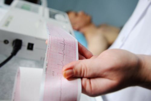
4. Notes when performing electrocardiogram

Lưu ý khi thực hiện điện tâm đồ
Doctor Nguyen Quoc Viet has more than 20 years of experience in the examination and treatment of cardiovascular diseases and Cardiovascular Interventions (Including angiography, dilation, stenting of coronary arteries, renal arteries...), placing temporary, permanent pacemaker... Before working at the Department of Medical Examination & Internal Medicine, Vinmec International General Hospital Da Nang, the doctor used to work for a long time at Da Nang Hospital.
Please dial HOTLINE for more information or register for an appointment HERE. Download MyVinmec app to make appointments faster and to manage your bookings easily.