This is an automatically translated article.
Posted by Doctor Mai Vien Phuong - Department of Examination & Internal Medicine, Vinmec Central Park International General Hospital
Hepatic hemangioma (HH) is the most common benign liver tumor and it is often discovered incidentally during physical examinations. This tumor arose from a vascular malformation; however, the pathophysiology has not yet been elucidated.
1. Concepts
Liver hemangioma presents as an incidental finding during abdominal ultrasound and is described as a solitary or multiple lesion. They may be limited to one lobe (more so in the right lobe) or extend to the entire liver. According to their size, they can be small or giant (>5 cm) and can be from 1 mm to 50 cm long. Liver hemangiomas are classified according to their nature as cavernous, capillary and sclerosing hemangiomas; The latter are characterized by degeneration and replacement of fibers and may be misdiagnosed as melanoma
2. Pathophysiology of liver hemangioma
The pathophysiology of hepatic hemangiomas is not completely understood, and in some cases a genetic predisposition has been described. Hemangiomas arise from a vascular malformation with a growth pattern secondary to dilation rather than hypertrophy or hyperplasia.
One hypothesis is that liver hemangiomas are the result of abnormal angiogenesis and an increase in pro-angiogenic factors.
Vascular endothelial growth factor (VEGF) is an important angiogenic factor for endothelial cells. The mammalian target rapamycin (mTOR) stimulates the auto-loop of VEGF signaling and cell proliferation in vascular endothelial cells. The TOR proteins are a class of serine/threanine kinases involved in ribosome biosynthesis, mRNA translation, and cell mass growth and proliferation. Zhang et al found increased expression of VEGF-A, pro-matrix metalloproteinase 2, and activated metalloproteinase 2 in HH cells compared with normal human liver endothelial cells.
Rapamycin inhibits mTOR and has been investigated in mouse and murine cell models as a possible treatment for vascular cell growth (mainly melanoma).
Rapamycin is currently used as an antifungal, antineoplastic, and antibacterial macrolide, but there are no human studies targeting liver hemangiomas.
Hormones like estrogen play a role in liver hemangioma growth, as they are seen more often in women and their size increases after hormone replacement therapy (HRT), oral contraceptives ( OCP) and pregnancy. The direct mechanisms of hormonal effects are known, such as liver hemangiomas are both estrogen and progesterone receptor negative and current evidence does not support a contraindication to OCPs/HRT/anabolic steroids in patients with liver hemangioma.
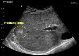
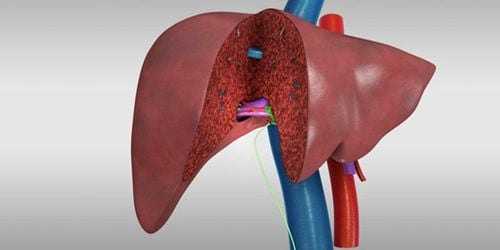
Hình ảnh u gan trên kết quả siêu âm
3. Symptoms of liver hemangioma
Liver hemangiomas are usually asymptomatic, however symptoms may appear when the HH is larger than > 5 cm. Symptoms are nonspecific, and patients often describe right upper quadrant abdominal pain, discomfort, and fullness, secondary to distension and Glisson's cyst. There is a tumor > 10 cm with abdominal distention. The location of the liver mass can put pressure on and compress adjacent structures causing other symptoms such as nausea, early satiety, and postprandial bloating. Less common symptoms include fever, jaundice, dyspnea, high-output heart failure, and serum sickness.
Giant liver hemangiomas can cause a life-threatening coagulation disorder known as Kasabach-Merrit syndrome (thrombocytopenia, disseminated intravascular coagulation, and systemic bleeding) with secondary coagulopathy. Occurs after thrombocytopenia, anemia, decreased fibrinogenimia, decreased prothrombin time and increased D-dimer. This syndrome has been reported with an incidence ranging from 0.3% of total HHs to 26% in tumors >15 cm [19, 25].
Another serious complication is bleeding due to spontaneous rupture or trauma (in giant peripheral and peripheral lesions), however the risk is extremely low (0.47%)
Currently, Vinmec International General Hospital has Hepatobiliary Screening packages, which help detect Hepatitis Virus at an early stage even when there are no symptoms. In addition, the comprehensive hepatobiliary screening package helps customers:
Evaluate the liver's ability to work through liver enzyme tests; Evaluation of bile function; vascular nutrition; Early screening for liver cancer; Perform tests such as Total blood cell analysis, blood clotting ability, screening for hepatitis B, C Assessment of liver and biliary status through ultrasound images and diseases that are at risk of affecting liver disease / liver disease. more severe liver disease In-depth analysis of parameters to evaluate hepatobiliary function through laboratory and subclinical tests; the risk of affecting the liver and early screening for hepatobiliary cancer.
Please dial HOTLINE for more information or register for an appointment HERE. Download MyVinmec app to make appointments faster and to manage your bookings easily.
ReferencesIshak KG, Rabin L. Benign tumors of the liver. Med Clin North Am . 1975;59:995-1013. [PubMed] [DOI] Yang Z, Tan H, Liu X, Sun Y. Extremely Giant Liver Hemangioma (50 cm) with Kasabach-Merritt Syndrome. J Gastrointest Surg . 2017;21:1748-1749. [PubMed] [DOI] Schumacker HB. Hemangioma of the liver: discussion of symptomatology and report of patient treated by operation. Surgery . 1942;11:209–222. Reddy KR, Kligerman S, Levi J, Livingstone A, Molina E, Franceschi D, Badalamenti S, Jeffers L, Tzakis A, Schiff ER. Benign and solid tumors of the liver: relationship to sex, age, size of tumors, and outcome. Am Surg . 2001;67:173-178. [PubMed] Hasan HY, Hinshaw JL, Borman EJ, Gegios A, Leverson G, Winslow ER. Assessing normal growth of hepatic hemangiomas during long-term follow-up. JAMA Surg . 2014;149:1266-1271. [PubMed] [DOI] Monica Leon, Luis Chavez, Salim Surani, Hepatic hemangioma: What internists need to know, World J Gastroenterol. Jan 7, 2020; 26(1): 11-20