This is an automatically translated article.
The article was professionally consulted by Specialist Doctor I Vo Thi Thuy Trang - Gastroenterologist, Department of Medical Examination & Internal Medicine - Vinmec Danang International Hospital. The doctor has many years of experience in the field of gastrointestinal endoscopy.The liver is a large organ that plays an important role in the digestive system. The liver is located in the peritoneum, located in the upper layer of the transverse mesentery, the right subdiaphragmatic cell but partially encroaching on the left inferior diaphragmatic and epigastric space. They have the function of detoxification, metabolism of glucide, protid, lipid..
1. Anatomical features of the liver
The liver is the largest organ in the body, belonging to the digestive system, accounting for 2% of body weight in adults and 5% in newborns. The liver has a smooth brown color when removed from the living body, easily broken, when ruptured or leads to submembrane haemorrhage (sub-membrane bleeding).In the human body, the liver weighs 1.4 - 1.8 kg in men and 1.2 - 1.4 in women, if counting 800 - 900ml of blood the liver contains, the average weight of the liver is 2.3 - 2.4 kg. The width of the liver is 25-28cm long, 16-20cm wide front and back, 6-8cm high (thick).
The liver has two faces: the convex diaphragm and the flat visceral surface. The posterior boundary is not clear, the front is the sharp edge called the lower border. Depending on the individual organ, the external appearance of the liver may vary slightly from normal.
2. Division of liver lobes according to anatomical landmarks
The superior surface of the liver (diaphragmatic surface) is divided into 2 lobes by the crescent ligament:Right lobe Left liver lobe
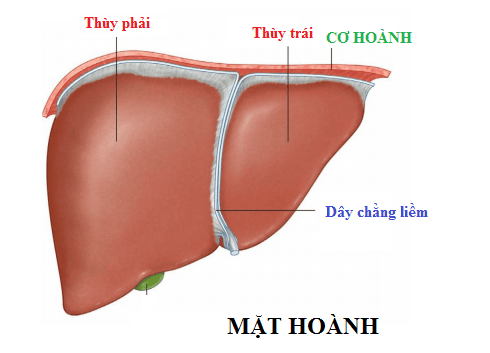
Hình ảnh mặt hoành
Right lobe of the liver Left lobe Square lobe Tail lobe.
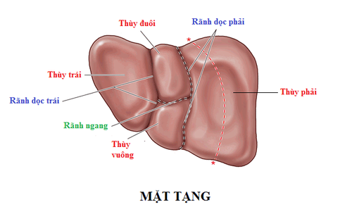
Hình ảnh mặt tạng
3. Division of liver lobes based on hepatic venous system according to Goldsmith and Woodburne
The hepatic vein divides the liver into 2 lobes (left lobe, right lobe) and 5 lobes (lateral lobe, caudal lobe, middle lobe, anterior lobe, posterior lobe) specifically as follows:The superior middle hepatic vein divides the liver divided into 2 parts: left and right The right superior hepatic vein divides the liver into 2 segments: anterior and posterior The left superior hepatic vein divides the liver into 2 segments: medial and lateral. The caudal lobe is independent, the hepatic vein drains blood directly into the inferior vena cava.
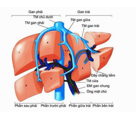
Phân chia thùy gan dựa vào hệ thống tĩnh mạch gan theo Goldsmith và Woodburne
4. Division of liver lobes according to Couinaud
Based on the 3-dimensional view of the distribution of the portal and hepatic veins, the liver is divided into 8 subsegments. The hepatic veins divide into 2 lobes (right and left hepatic lobes) and 5 lobes (tail, lateral, medial, anterior, posterior).The right and left portal veins divide the segments into superior and inferior subsegments, except for the middle segment. The lobes are numbered clockwise If viewed from the anterior view, the order from I is caudal subsegment VIII.
5. Liver lobe division according to Professor Ton That Tung
The liver has 2 lobes, left and right, and has 5 lobes: anterior, posterior, medial, lateral, and dorsal. The main dorsal segment is the caudal lobe or the lower segment I. The liver has only 6 lower segments: II, III, V, VI, VII, VIII. Thus, the middle segment, also known as the square lobe, is the lower segment IV.The numbering of the subsegments from I to VIII according to the division of Couinaud's author is not as well applied in liver resection as GS's. Ton That Tung.
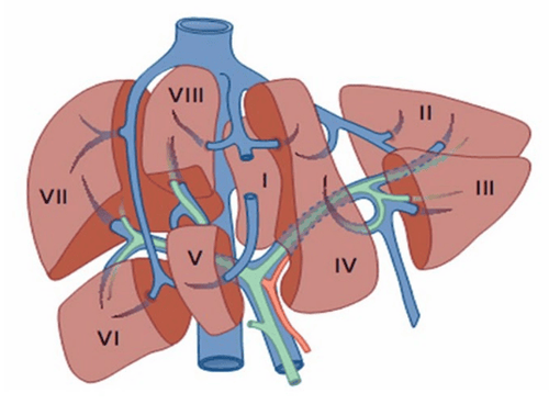
Sự đánh số các hạ phân thuỳ từ I đến VIII theo cách phân chia của tác giả Couinaud
Segment The left side of the liver is divided by the left branch of the portal vein into inferior segment II and inferior segment III. The anterior segment of the right liver, divided by the right branch of the portal vein, into inferior segment VIII and inferior segment V. The right posterior segment of the liver is divided by the right branch of the portal vein into inferior segment VII and inferior segment VI. The caudal lobe is called the lower lobe I. The middle lobe of the left liver is called the lower segment IV.
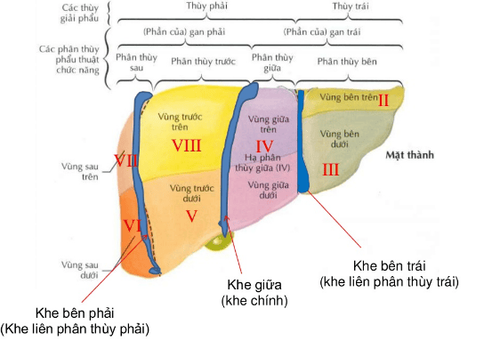
Hình ảnh nhìn từ mặt hoành
Midhepatic fissure: On the diaphragmatic side of the liver, the midhepatic cleft goes from the cyst of the gallbladder to the left border of the inferior vena cava; on the visceral surface, the midhepatic cleft goes from the middle of the gallbladder fossa to the left border of the inferior vena cava. . The midhepatic slit divides the liver into two halves, the right and the left, and in the midhepatic fissure contains the middle hepatic vein. The right intersegmental cleft is defined from the right border of the inferior vena cava, parallel to the right border of the liver, three fingers from the right border of the liver, which contains the right hepatic vein and divides the right liver into two segments. are the posterior and anterior segments. Left intersegmental fissure: On the diaphragmatic surface, the left intersegmental fissure is the attachment line for the crescent ligament, on the visceral surface the left intersegmental fissure corresponds to the left longitudinal fissure and contains the left hepatic vein. This fissure divides the left liver into two segments, the medial and the lateral. The right midlobular fissure is not obvious: The midlobe fissure divides the anterior segment into subsegments V and VIII, and the posterior segment into subsegments VI and VII. Left middle lobe accessory fissure: On the diaphragmatic plane, the left middle lobe accessory fissure goes from the left border of the inferior vena cava to the posterior 1/3 and anterior 2/3 of the lower border of the left liver, on the visceral surface this fissure will go from the left end of the left lobe. The liver connects the posterior 1/3 and anterior 2/3 of the lower border of the left liver, divides the lateral segment into segments II and III, and the lower segment I corresponds to the caudal lobe of the liver. Knowing how to divide liver lobes is very important, so that doctors can diagnose and offer treatment methods for some hepatobiliary diseases.
Vinmec International General Hospital is the address for examination, treatment and prevention of diseases, including Gastroenterology. When performing the examination process at Vinmec, customers will be welcomed and used the facilities and modern machinery system along with perfect medical services under the guidance and advice of the doctors. Good doctors, well-trained both at home and abroad.
Please dial HOTLINE for more information or register for an appointment HERE. Download MyVinmec app to make appointments faster and to manage your bookings easily.