This is an automatically translated article.
Posted by Master, Doctor Mai Vien Phuong - Department of Examination & Internal Medicine - Vinmec Central Park International General HospitalThe pathogenesis of calcified fibroblastomas remains unclear, but some have hypothesized that they may be involved in IgG4-related disease, inflammatory fibroblast injury, and vascular Castleman disease. hyalinemia, sclerotic nodular changes of the spleen, or trauma.
1. Pathogenesis of calcified fibrous tumors
Currently, the pathogenesis of calcified fibrosarcoma is uncertain. Etiology may include infection, trauma, or previous surgical intervention. Plasma cells in calcified fibroblasts may stain positively for IgG and IgG4, which raises the possibility that calcified fibroblasts may be indicative of IgG4-associated disease (IgG4-RD). . IgG4-RD leads to the formation of pseudotumors, most commonly in the pancreas. Similar to calcified fibrosarcoma, IgG4-RD gives rise to clinically benign inflammatory mass lesions. However, IgG4-RD giving rise to mass lesions in the stomach is rare, although such cases have been reported.Histologically, IgG4-RD showed storiform fibrosis, lymphocytic inflammation, and obstructive phlebitis. Similarly, calcified fibroblasts show lymphocytic tissue infiltration, and dense fibrosis seen in calcified fibromas often shows a vague but non-classical pattern. Cases of calcified fibroma tumors with IgG4-positive plasma cytopenia have been reported, sometimes with elevations in serum IgG4. However, cases of calcified fibrosarcoma in patients with other manifestations of IgG4-RD such as type I autoimmune pancreatitis are often missing. Histological evidence of obstructive phlebitis, a key endpoint of IgG4-RD, has not been conclusively demonstrated in calcified fibrous tumors. Furthermore, the detection of differentially elevated IgG4-positive plasma cells is nonspecific, and can be seen in many inflammatory conditions.
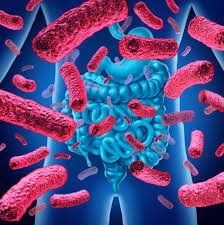
Nhiễm trùng có nguy cơ gây ra u xơ hóa vôi hóa
2. It is hypothesized that calcified fibroblast tumors may represent the late sclerotic stage of inflammatory fibroblast tumors:
It has been hypothesized that calcified fibroblastic tumors may represent the late sclerotic stage of inflammatory fibroblast tumors (IMT). This hypothesis was formed by the observation that calcified fibrosarcoma and IMT can coexist in close proximity in the same patient. Late-stage IMTs are cells that form with dense fibrosis and are sometimes associated with calcification. Studies showing the presence of ALK rearrangements in IMT, which are absent in calcified fibrosarcomas, have suggested that calcified fibrosarcoma is a distinct neoplastic process. However, genome-wide methylation analysis revealed overlapping methylation patterns of calcified fibrosarcoma and IMT, suggesting that both lesions may represent a spectrum of the same disease, regardless of gene fusion analysis.3. Calcified fibrous tumor is also associated with a number of other diseases
Calcified fibrous tumors have also been associated with hyaline vascular type Castleman disease, although such an association appears to be rare. Castleman disease is a benign lymphoproliferative disorder that can occur in a single lymph node (monocentric) or multiple lymph nodes (multicentre). The hyalinized vascular type is characterized by germinal centers surrounded by concentric rings of mantle lymphocytes and interfollicular hyalinized vascular proliferation. Six cases were reported between 1999 and 2019, in which calcified fibrosarcoma was found to be associated with hyaline vascular type Castleman disease. In some cases, both calcified fibrosarcoma and hyaline vascular type Castleman disease were found to coexist in the same lymph node tissue, while in other cases, calcified fibrous tumor patients gastrointestinal tract was found to have hyaline vascular type Castleman disease in the adjacent lymph nodes. The etiology of this association remains unclear and raises the possibility that both entities may represent different stages of the same reactive disease process. It has been suggested that calcified fibrous tumor-like features in the lymph nodes of patients with hyaline vascular type Castleman disease are the result of fine needle aspiration trauma, supporting the view that calcified fibrous tumors may be a proliferative response to trauma. Further supporting this theory are reports of patients who developed soft tissue calcified fibrosarcomas within a few months of trauma maintained in the same anatomical location.Trauma hypothesis:
However, many cases of calcified fibrous tumors in the GI tract were not associated with a history of trauma or acute tissue trauma such as peptic ulcer or perforation, suggesting a calcified fibrous tumor of the GI tract. may result from chronic tissue injury, rather than acute trauma, or may have an entirely different pathogenesis. Passage of food can cause localized tissue damage in the gastrointestinal tract, which can trigger a sclerotic inflammatory response, which eventually gives rise to calcified fibrous tumors and is likely to account for the relatively high frequency of cystic fibrosis. calcified fibrous tumors in the gastrointestinal tract.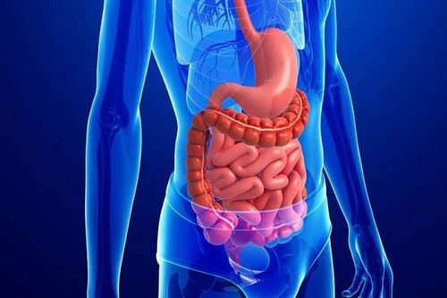
Thức ăn có thể gây tổn thương đường tiêu hóa dẫn đến u xơ hóa vôi hóa
The relationship between calcified fibrous tumor and sclerotic nodular change of the spleen:
Another rare association has been made between calcified fibrosarcoma and sclerosing vascular nodular transformation of the spleen (SANT). SANT refers to non-tumor vascular proliferation in the spleen of unknown cause, histologically characterized by nodules with vascular spaces lined with plump endothelial cells interspersed with ovoid cells. or rhombus, surrounded by concentric collagen fibers. Rare cases of SANT coexisting with diffuse intra-abdominal calcified fibrosarcoma have been described, and suggest a possibly common response mechanism. In addition, increased IgG4-positive plasma cells were observed in SANT and in associated calcified fibrous tumors, suggesting that both processes may be associated with IgG4 pathophysiology. -RDThe pathogenesis of calcified fibrous tumors remains unclear
Finally, the pathophysiology of calcified fibrous tumors remains unclear, although emerging evidence suggests that these lesions may be pathophysiologically related to other inflammatory lesions such as IgG4-RD. , IMT, hyaline vascular Castleman disease, or SANT. Calcifying fibroma may represent a late stage manifestation of any one of these lesions. Alternatively, calcified fibroma may simply represent a late stage of a variety of inflammatory and sclerotic processes, without regard to a specific etiology. This may account for the association of calcified fibroblasts with many other types of lesions. Other genetic and/or environmental factors, such as trauma, may contribute to calcified fibroma, and the aetiology may vary by anatomical site. More studies are needed to further elucidate the pathogenesis of calcified fibrous tumors.Please dial HOTLINE for more information or register for an appointment HERE. Download MyVinmec app to make appointments faster and to manage your bookings easily.
References
Turbiville D, Zhang X. Calcified fibrous tumors of the gastrointestinal tract: Clinical pathology review and update. World J Gastroenterol 2020; 26 (37): 5597-5605 [PMID: 33071524 DOI: 10.3748 / wjg.v26.i37.5597 ]