This is an automatically translated article.
The article was professionally consulted by Specialist Doctor I Le Nguyen Hong Tram - Gastroenterologist - Department of Medical Examination & Internal Medicine - Vinmec Nha Trang International General Hospital.Fatty liver ultrasound and liver tissue elastography have good effects in detecting fatty liver disease early. This method adopts images that accurately reflect the liver condition with relatively high reliability.
1. Overview
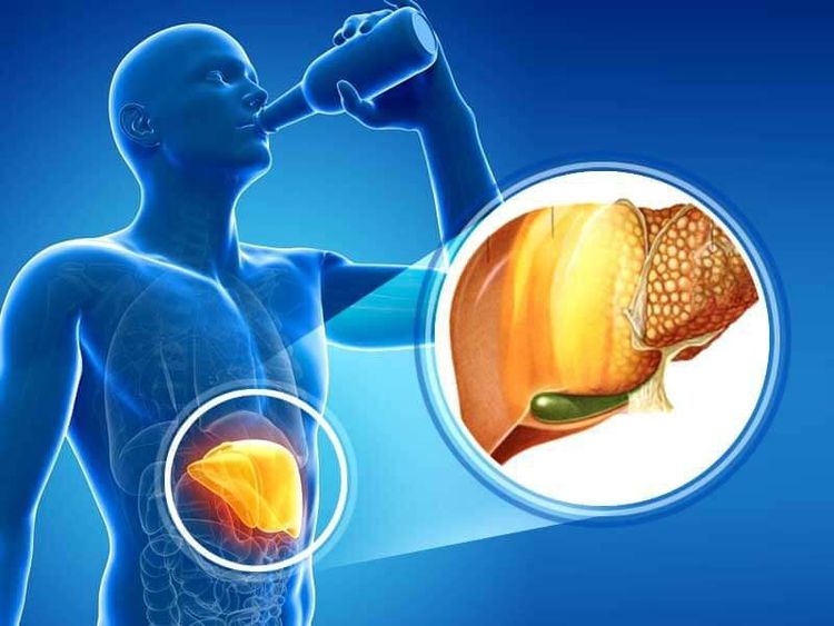
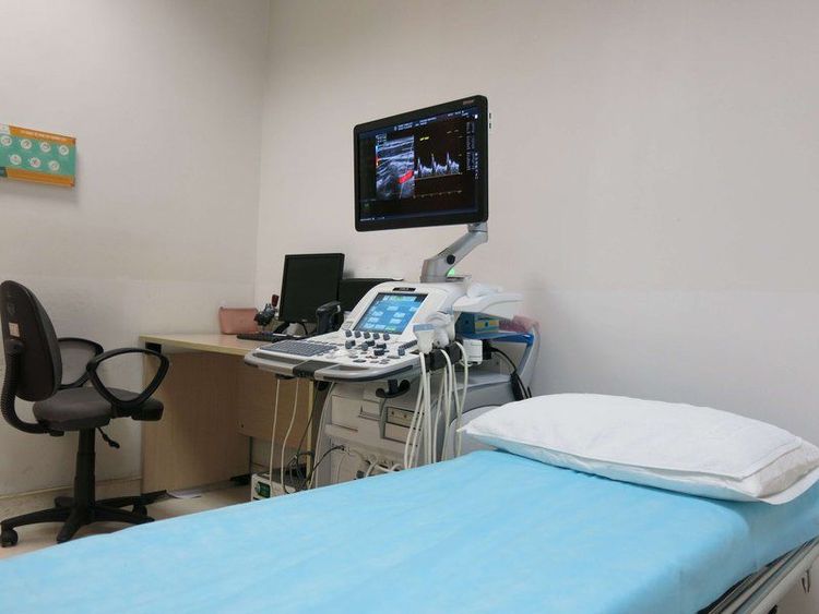
The liver is the second largest organ in the human body (average 1.44kg - 1.66kg), located in the upper right side of the abdomen. The liver's role is to process virtually everything we eat or drink, while also removing toxins from the body, producing proteins, and producing bile needed for digestion. Normally, fat in the liver is normal, but too much fat in the liver is called fatty liver disease, which affects the functioning and function of the liverFatty liver ultrasound is the diagnostic method used High-frequency sound waves hit body areas near the liver to create images of the internal organs. During the ultrasound, the doctor will use an ultrasound machine with an instrument that sends ultrasound waves to the upper right side of the patient's abdomen to see images of the liver. These images will be displayed through a computer screen, helping to detect abnormalities in the liver structure and severe damage to the liver.
2. Classification of fatty liver
Fatty liver is divided into 4 main groups of diseases:Non-alcoholic fatty liver (NAFLD): caused by disorders of fat metabolism of the liver, leading to excess fat in the liver's organs. Patients are considered to have NAFLD when the percentage of fat in the liver accounts for more than 10% of the weight of the liver. Alcoholic fatty liver: Excessive alcohol consumption will damage the liver and lead to impaired fat metabolism. Abstinence from alcohol helps to reduce fatty liver. After 6 weeks, your liver will no longer be fatty. An important note here is that excessive and continuous alcohol use increases the risk of fatty liver disease progressing to cirrhosis. Non-alcoholic steatohepatitis (NASH): The cause of fatty liver is not due to alcohol, but because the amount of fat in the liver reaches a certain level, the liver enlarges and may be accompanied by a decline in liver function. The main symptoms include: loss of appetite, vomiting, nausea, abdominal pain, jaundice. If the disease is not treated, it will lead to irreversible damage to the liver and eventually cirrhosis. Acute fatty liver during pregnancy: is a rare complication and if left untreated, can endanger the life of the pregnant woman. Symptoms appear in the last 3 months of pregnancy, including: persistent nausea and vomiting, pain in the right lower quadrant, jaundice, and discomfort throughout the body. Pregnant women will be screened and prevented early through ultrasound of fatty liver and ultrasound of liver tissue elasticity. Most symptoms will subside after delivery and leave no consequences later.

3. Diagnosis of fatty liver
Fatty liver disease is often discovered during a general examination. The first sign of the disease is usually from the results of an imaging test. In addition, the doctor may order the following methods and tests:Blood tests are used to diagnose viral hepatitis (hepatitis A, B, or C) Liver function tests to assessment of liver function and concentrations of substances produced or metabolised in the liver. With the method of elastography of liver tissue, the typical image of fatty liver will appear with the liver parenchyma "bright" and thicker, the liver is usually larger and the liver is round and smooth (light liver). The method of ultrasound for fatty liver has a sensitivity of over 90% and is currently widely used. Besides, there are a number of other imaging modalities such as computed tomography or magnetic resonance imaging. Although imaging helps detect fatty liver, it does not fully assess liver function and many other lesions. Perform a liver biopsy to make a more accurate diagnosis. Liver biopsy is a medical technique that uses a biopsy needle to remove a piece of liver tissue and send it for cytological examination. This is considered a good method to diagnose as well as determine the cause of fatty liver.

4. Fatty liver ultrasound results
4.1 Description of fatty liver on ultrasound
When ultrasound of fatty liver, if there are abnormal signs, on the liver morphology image, there will appear scattered light spots or concentrated in each area. Depending on the degree of liver fat in each subject, this condition will have different degrees. Also through ultrasound, the doctor will not be able to see or clearly see the external vascular system on the liver.Perform a venous examination of the liver, portal vein or diaphragm and the doctor finds no echoes. Besides, the echo of the machine will also be reduced at the back of the liver. Thus, images of the margins of the liver and liver become clear. In general, the ultrasound depiction of fatty liver is characterized by increased echogenicity of the liver parenchyma resulting in a characteristic bright liver image.
4.2 Evaluation of fatty liver results by ultrasound
If fatty liver ultrasound shows part or all of the liver parenchyma bright enough to determine the extent of the disease. Accordingly, based on the phenomenon of increased brightness of the liver parenchyma on the ultrasound image, the degree of fatty liver is assessed as follows:Level 1: feel a slight increase in the diffuse echo of the parenchyma. , the level of sound absorption has not changed significantly and the visibility of the diaphragm region as well as the venous border is still quite clear. Level 2: The diffuse echo and echogenicity of the parenchyma is increased, the diaphragm can be identified, but the visibility of the diaphragm and the intrahepatic venous border is more blurred than before. Grade 3: There is a marked increase in diffuse echogenicity and parenchymal aspiration, so high that the diaphragm region and the intrahepatic venous border are no longer visible. These ultrasound descriptions of fatty liver will help the doctor determine the extent of the disease and give the right treatment plan for each patient.
Vinmec International General Hospital is implementing the Hepatobiliary Screening Package to help diagnose fatty liver disease in particular and hepatobiliary diseases in general. Vinmec is a prestigious and reliable address in the field of screening and treatment of liver and biliary diseases, with the following advantages:
Possessing modern facilities, full of world advanced equipment and machinery. , for high image quality, accurate and fast test results, maximum support for disease diagnosis and treatment. A team of specialized medical doctors from major hospitals across the country helps the treatment process to be highly effective and shorten the treatment time. Professional, comprehensive examination and consultation service, civilized, polite, safe and sterilized medical examination space. Specialist I Le Nguyen Hong Tram graduated as a Doctor of General Internal Medicine at the University of Medicine and Pharmacy in Ho Chi Minh City. Ho Chi Minh, doctor has many years of experience in the examination and treatment of gastrointestinal diseases, especially gastrointestinal - hepatobiliary diseases, gastrointestinal endoscopy and general internal diseases. Currently, Dr. Tram is working at the Department of Medical Examination and Internal Medicine, Vinmec Nha Trang International General Hospital.
Please dial HOTLINE for more information or register for an appointment HERE. Download MyVinmec app to make appointments faster and to manage your bookings easily.


