This is an automatically translated article.
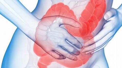
Posted by Master, Doctor Mai Vien Phuong - Department of Examination & Internal Medicine - Vinmec Central Park International General HospitalMicroscopic colitis is a chronic inflammatory bowel disease with chronic diarrhea symptoms but the patient has a normal colonoscopy. Diagnosis of microscopic colitis is based on histopathology.
1. History of microscopic colitis
Microscopic colitis is divided into two groups, namely collagenous colitis and lymphocytic colitis.
Collagen colitis was first described in 1976, in a patient with chronic diarrhea who had a biopsy of the rectal mucosa. Biopsy results showed dense collagen deposition in the subepithelial layer. In 1980, Read et al. first used the term "microscopic colitis" to describe a disease characterized by chronic diarrhea and colorless stools. Then, in 1989, Lazenby et al. described the clinical and histopathological features of lymphoid colitis. These two diseases have many similarities in terms of clinical and epidemiological features, so they are grouped together in the group of microscopic colitis.
According to one study, the annual incidence of collagenous colitis and lymphocytic colitis per 100,000 people was 4.14 and 4.85, respectively. The prevalence rates of collagenous colitis and lymphoid colitis were 49.21 and 63.05 per 100,000 people, respectively. The incidence of these two forms of colitis varies by region, but mainly in Europe and America. In the Asian region, a number of studies in Korea recorded the rate of collagenous colitis in patients with chronic diarrhea at 4%, similar to the rate in Western countries but higher than in the study. in Malaysia and India.
2. Risk factors for microscopic colitis
2.1. Autoimmune diseases There is an association between microscopic colitis and autoimmune diseases such as Celiac disease, polyarthritis, thyroid disease. There are 20 - 60% of patients with lymphocytic colitis and 17 - 40% of patients with collagenous colitis have concomitant autoimmune diseases. According to one study, up to 30% of Celiac patients present with microscopic colitis. When compared with rates in the community, Celiac patients had a 70-fold higher rate of progression to microscopic colitis, of which 64% were found after the diagnosis of Celiac, 11% were diagnosed before. and 25% diagnosed at the same time.
2.2. Smoking In a study conducted from 2007 to 2010 in Spain on 255 smoking patients showed a strong association with lymphocytic colitis and collagenous colitis with OR 3, respectively. ,8 and 2.4.
Another Swedish study also found that smoking was associated with collagenous colitis and that smokers developed the disease 14 years earlier than non-smokers. The hypothesis proposed to explain this association is that smoking reduces the blood flow to the colon, thereby altering the permeability of the colonic mucosa. 2.3. Use of certain drugs Beaugeri et al.'s research has found an association of several drugs with the occurrence of microscopic colitis. A few reports have also reported that patients with microscopic colitis improved their clinical symptoms when NSAIDs were discontinued and relapsed upon re-initiation.
In addition, concomitant use of proton pump inhibitors (PPIs) and NSAIDs may increase the risk of developing microscopic colitis. Of the drugs in these two classes, diclofenac and omeprazole have been the most commonly reported.
2.4. Other factors Microscopic colitis is more common in women, however a Swedish study found no association between sex hormone levels and microscopic colitis. Several studies have addressed the role of bile acid malabsorption in microscopic colitis. Histopathological results noted villi atrophy, inflammation and collagen deposition in the ileum in cases of bile acid malabsorption. A Spanish study reported that 43% of patients with microscopic colitis had bile acid malabsorption and were more common in lymphoid colitis.

3. Pathogenesis
The pathogenesis of microscopic colitis is still unclear, but several cohort studies have hypothesized to be related to autoimmune or immune dysregulation during exposure. exposure to antigens, which are certain drugs or microorganisms. Exposure to antigens is associated with epithelial barrier dysfunction that alters the permeability of the epithelial layer, and antigens can act on the subepithelial layer. The overlap with Celiac in the expression of HLA genes suggests the hypothesis that alterations in antigen presentation occur in the binding stroma of the mucosa. To date, there are still relatively few data on the role of the innate immune response, and most of them focus on describing the abnormality of the protective barrier in the intestinal mucosa. Increased concentrations of nitric oxide in the colonic mucosa in microscopic colitis will affect the junction between epithelial cells, thereby leading to increased permeability in this area. This phenomenon is also seen in a number of other inflammatory diseases of the intestinal tract and is considered to play an important role in the pathogenesis.4. Mechanism of diarrhea in microscopic colitis
To date, several hypotheses have been proposed to explain the mechanism of diarrhea in microscopic colitis, including:
Change in mucosal permeability due to inflammatory process. Polymorphisms of serotonin reuptake transporters affect intestinal mucosal motility and secretion. Bile acid malabsorption. Inflammatory mediators in the epithelial chorion are caused by antigens such as drugs, food, bile salts, bacteria that activate lymphocytes.
5. Clinical symptoms of microscopic colitis
Clinically, it is indistinguishable between collagenous colitis and lymphocytic colitis. The prominent symptom of microscopic colitis is chronic diarrhea but no blood in the stool, the patient has a normal gastrointestinal endoscopy picture.
Chronic diarrhea is defined as when the condition persists for more than 4 weeks. The stools are watery, which causes the patient to feel the need to defecate immediately (about 70% of patients) or to fecal incontinence (about 40% of patients). In severe cases, patients may have more than 15 bowel movements/day and often have nocturnal diarrhea (50% of patients). However, severe dehydration, electrolyte disturbances, or other complications are uncommon. In advanced stages of the disease, patients often have a significant impact on quality of life. Abdominal pain is also a common symptom. The incidence of discomfort or abdominal cramps can be as high as 50%. The differential diagnosis between microscopic colitis and irritable bowel syndrome is difficult.
Besides, microscopic colitis is also common in autoimmune diseases. According to a multicenter study in Sweden, the prevalence of autoimmune diseases was found in 1/3 of patients with microscopic colitis, which included Celiac disease (12%), autoimmune thyroiditis (10.3 %) %), Sjogren's syndrome (3.4%), diabetes mellitus (1.7%), autoimmune lesions of the skin and joints (6%). In most cases, autoimmune disease is diagnosed before microscopic colitis.

6. Subclinical diagnosis
Diagnosis of microscopic colitis is usually based on clinical symptoms, histopathological findings and exclusion of other pathologies based on endoscopic images as well as other imaging methods normal. Tests of patients with microscopic colitis in most cases are normal or have nonspecific changes such as mild elevation of C-reactive protein, mild anemia. The definition of microscopic colitis is inflammation of the colon that is diagnosed by histopathology and the patient has a normal total gastrointestinal endoscopy result, so a total colonoscopy is the exploratory step. important in disease diagnosis.
When performing endoscopy, attention should be paid to detecting colonic mucosal lesions as well as multi-segment biopsies in colon segments. A 2-piece biopsy of each segment of the colon is recommended for a more accurate histopathological diagnosis, and biopsies of all segments are more useful than focusing on only one segment. Gastrointestinal findings may be normal or nearly normal. With the development of endoscopic techniques, mucosal lesions in microscopic colitis have been reported and evaluated as supporting evidence for the diagnosis.
A study on patients with collagenous colitis in Japan reported that up to 80% of patients had lesions on endoscopy, including: vascular proliferation, unclear mucosal vascular morphology, longitudinal ulcers, oil "cat-scratch" sign, regional edema, mucosal papules.... Longitudinal mucosal lesions have been reported in patients with lansoprazole-associated collagenous colitis. There have been case reports of collagenous colitis with severe mucosal damage and complications.
7. Approach to diagnosis of microscopic colitis
Take the history:
Take the characteristics of diarrhea, blood mucus, frequency, course, time to progress, nocturnal diarrhea, fecal incontinence. Skinny weight loss. Family history (IBD, Calia, multiple endocrine malignancies). Drug history (abuse of laxatives, adverse drug reactions, radiation enteritis, gastrointestinal surgery, cholecystectomy). Systemic symptoms:
Diet (lactose malabsorption, fructosa, sorbitol, Celiac). Occupational history, travel, HIV exposure, persistent recurrent infections. Diet (lactose malabsorption, fructosa, sorbitol, Celiac). Assess the patient with the following tests:
Stool culture: Clostridium difficile, stool culture, fresh parasitoscopy, Giardia fecal antigen. Serum tests: Blood count, red electrolytes, protein, albumin, blood pressure, thyroid function. Serum test for Celiac disease. Test for lactose intolerance. Endoscopy and biopsy. Histopathology.

8. Treatment of microscopic colitis
Treatment of microscopic colitis depends on the severity of symptoms. The goal of treatment is to reverse the disease and improve the patient's quality of life. In the case of patients with frequent relapses, maintaining a response to treatment is essential.
8.1. Avoiding Risk Factors This is the first step in the management of microscopic colitis. The relationship between smoking and use of certain medications should be discussed with the patient regarding the onset and progression of the disease.
8.2. Using Budesonide Budesonide is the only drug effective in controlled, clinical trials. Budesonide is a corticosteroid with local action, the drug is metabolized by the liver, has little systemic effect, so it is very safe.
8.3. Some Other Treatment Options Probiotics: Of the clinical trials involving the efficacy of probiotics for microscopic colitis, only one clinical trial using Boswellia serrata demonstrated an improvement in efficacy. clinical after 6 weeks. Antidiarrheal: Several studies have shown the effectiveness of loperamide in improving clinical symptoms of the disease, however, there are no studies demonstrating its effectiveness in reversing the disease with maintenance therapy. . Mesalazine: In a clinical trial in patients with microscopic colitis, masala zone 2.4 g/day with or without cholestyramine 4 g/day was maintained for 6 months. The rate of clinical remission and histopathological improvement was 85% in patients with lymphocytic colitis, 91% in patients with collagenous colitis, especially in the combination treatment group. Immunomodulators: Azathioprine and 6-mercaptopurine are used in patients with very severe symptoms who are unresponsive or intolerant to budesonide. Have a reasonable diet: In the nutritional diet, people with colitis should avoid eating hot spicy foods, foods that are not hygienic, fast foods, foods containing many protective chemicals. management, .... However, many people also wonder about the issue of "can you eat yogurt with colitis or can you drink milk with colitis?". In fact, people with microscopic colitis do not need to avoid eating yogurt, as it provides a good amount of probiotic bacteria, helping the intestines absorb nutrients better. In summary, microscopic colitis has clinical symptoms such as chronic diarrhea, no blood, watery stools. The disease is of autoimmune origin, the etiology is still unclear. Therefore, the main goal of treatment is to eliminate the factors that aggravate the disease and control the symptoms.
Please dial HOTLINE for more information or register for an appointment HERE. Download MyVinmec app to make appointments faster and to manage your bookings easily.
References:1. Dao Van Long, Dao Viet Hang. Autoimmune diseases of the gastrointestinal tract. Medical Publishing House.
2. Lindstrom C.G. (1976). “Collagenous colitis” with watery diarrhea--a new entity? Pathol Eur, 11(1), 87-89.
3. Read N.W., Krejs G.J., Read M.G. et al. (1980). Chronic diarrhea of unknown origin. Gastroenterology, 78(2),264-271.
4. Lazenby A. J., Yardley J.H., Giardiello F. M. et al. (1989) Lymphocytic ("microscopic") colitis: a comparative histopathologic study with particular reference to collagenous colitis. Pathol, 20(1), 18-28.


