This is an automatically translated article.
This article is professionally consulted by Dr. Pham Quoc Thanh - Radiologist - Department of Diagnostic Imaging - Vinmec Hai Phong International General Hospital.1. What is magnetic resonance imaging (MRI)?
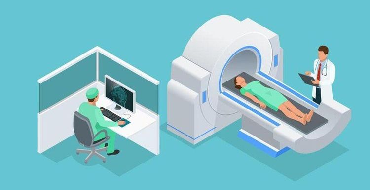
Magnetic resonance is a specialized tomography technique that uses magnetic fields and radio waves. Then, the hydrogen atoms in the patient's body, under the influence of magnetic fields, radio waves, will quickly release RF energy. And different body tissues will absorb and release different energy.The entire process of releasing this energy source will be received by the computer, processed, and converted into different image signals.
Images from MRI results have relatively high contrast, good anatomical details, and perfect 3D reconstruction, and do not bring side effects like X-rays, so present Nowadays, MRI is increasingly widely used for many different fields, for example: neurological, musculoskeletal, abdominal, cardiovascular,...
MRI is evaluated as one of the current imaging techniques. The greatest, revolutionary technology in medicine. To date, magnetic resonance imaging (MRI) is increasingly known for its safety, accuracy and non-invasiveness, without the use of X-rays. With its very high resolution images, and investigative capabilities. Multi-section will give the sharpest images of the location to be taken, and at the same time provide an assessment of the properties of the tissue under investigation.
2. Magnetic resonance imaging procedure of lower extremities without injection of contrast agent
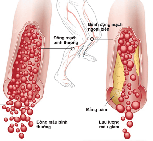
Magnetic resonance angiography of the lower extremities will provide important assessments of diseases involving the arteries of the lower extremities. This is a non-invasive method and does not use X-rays, so it does not cause health effects to the patient, this technique is increasingly popular, used to diagnose and monitor dynamic diseases. lower extremity circuit.2.1 When is non-injection magnetic resonance imaging of lower extremities indicated and contraindicated? MRI technique is indicated when the patient is experiencing arterial diseases of the lower extremities such as stenosis, aneurysm, occlusion, malformation or dissection, ... Or is also used to monitor after the treatment process, or at the request of specialist doctors. Absolute contraindications for the following cases: Patients carrying electronic devices, eg: defibrillators, pacemakers, automatic drug pumps under the skin, cochlear implants, ... Patients who are using surgical forceps made from metal in endoscopic, facial cavity or vascular < 6 months.


Magnetic resonance angiography of the lower extremities plays an important role in the analysis and evaluation of lower limb pathologies. This is an advanced, modern method that requires high skills, so you should go to a reputable medical facility to be examined and consulted with related questions.
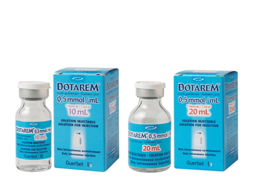
Vinmec International General Hospital is a unit that has a 3.0 Tesla magnetic resonance imaging machine system with modern Silent technology that brings outstanding advantages and owns an imaging room that meets the standards of the Ministry of Health. Accompanied by only the use of contrast agents very rarely cause adverse reactions in the patient. The scan time is about 45 - 60 minutes and the patient will receive the MRI results after 15-30 minutes, depending on the abnormalities in the body of the person taking the scan
3.0 Tesla Magnetic Resonance Imaging System with Silent technology of GE Healthcare (USA) is modernly equipped in hospitals. As a modern machine, high magnetic force, fast capture should evaluate blood vessels, flow clearly. And have the best image processing software. A team of experts from many different specialties will consult to provide the best diagnosis and treatment for the disease. For the 3.0 Tesla magnetic resonance imaging machine with Silent technology from GE Healthcare (USA) brings the Outstanding advantages such as:
Silent technology is especially beneficial for patients who are children, the elderly, weak health patients and patients undergoing surgery. reduce stress for the client during the shooting process, resulting in better image quality and shorter shooting time. Magnetic resonance imaging technology is the technology applied in the most popular and safest imaging method today because of its accuracy, non-invasiveness and no use of X-rays. Doctor. Pham Quoc Thanh has received intensive training and participated in many national and international scientific conferences on diagnostic imaging. The doctor has 13 years of experience in the field of diagnostic imaging and is currently a doctor at the Department of Diagnostic Imaging, Vinmec Hai Phong International General Hospital.
Please dial HOTLINE for more information or register for an appointment HERE. Download MyVinmec app to make appointments faster and to manage your bookings easily.


