This is an automatically translated article.
The article is professionally consulted by Dr., Doctor Tran Nhu Tu - Head of Diagnostic Imaging Department - Vinmec Danang International General Hospital.
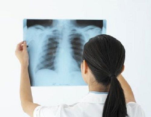
X-ray is a commonly used method in diagnostic imaging. However, many pregnant women are still very hesitant to take X-rays during pregnancy due to concerns that X-rays may adversely affect the baby. So specifically, how does the X-ray emitted from the X-ray technique affect the fetus?
1. Possible risks when taking X-rays
Possible risks after an X-ray are very rare. However, if taken many times, taking pictures over and over in a short time will have many potential health risks because X-rays can cause damage to some cells in the body and can progress to cancer. cancer later.
In order to obtain the clearest image of the organ to be taken as well as to ensure the health of the photographer, the dose of X-ray radiation is always kept at the optimal level.
Pregnant women should not have X-rays unless absolutely necessary because X-rays can cause abnormalities in the fetus. Usually, before taking an X-ray, doctors will ask if you are pregnant before deciding to perform this technique.
2. Effects of X-rays on the fetus
2.1 Mechanism of impact of X-rays on the fetus The influence of X-rays on human health is related to the dose of X-rays, exposure time, number of times of receiving X-rays.... But everyone needs to understand that Every day we still receive radiation from around, no one can avoid it.
X-rays have a lower dose than the radiation used for treatment. Therefore, the level of danger when exposed to X-rays also varies.
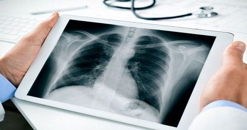
When performing X-ray techniques on organs such as the heart and lungs, X-rays do not shine into the fetal area. Some secondary rays can be reached, but in very small doses, it is unlikely to increase the risk of birth defects in the fetus.
According to studies, if the fetus is exposed to radiation doses of 2-6 rads, there is a risk of cancer later on, with radiation doses of 5-6 rads, the fetus can be at risk of malformations. natural.
X-rays in the diagnosis hardly increase the rate of birth defects in the fetus, but it is necessary to minimize the exposure to X-rays during pregnancy. When an X-ray is indicated, it is necessary to inform the doctor about your pregnancy.
2.2 The degree of influence of X-rays on the fetus Specifically, with the same radiation dose, the level of danger of X-rays to the fetus depends on each gestational age:
From 0-1 gestational week: X-rays can kill embryos From 2-7 weeks of pregnancy: X-rays can cause deformities, fetal growth retardation, cancer risk From 8-40 weeks: X-rays can cause deformities, fetal growth retardation, delay and risk of cancer For each imaging technique (X-ray, CT scan) in different organs, the rate of fetal damage will also be different with the dose Different rays:
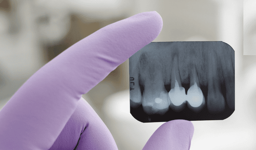
X-ray of abdomen, pelvis, pelvis, CT abdomen, chest: the rate of fetal damage is 1/100 000-1/10 000 (dose from 0.1-1) X-ray head, chest, neck CT scan, head: fetal injury rate is <1/1000 000 (dose 0.001-0.0001) X-ray of spine, lumbar, CT pelvis: rate of fetal injury from 1/10 000 to 1/1000 (dose 1-10) The fetus is very little affected by X-rays in the first 2 weeks. At much higher doses than 50 millisieverts, the new X-ray has the potential to cause miscarriage, which is equivalent to 500 cardiopulmonary scans. 2.3 Fetal X-ray dose in radiography Fetuses from 2 to 8 weeks: with diagnostic doses, X-rays are unlikely to cause malformations, miscarriages or slow fetal growth, except in Doses above 200 millisieverts are equivalent to 2000 cardiopulmonary scans. Fetal from 8 to 15 weeks: At this time, the fetal central nervous system may be sensitive to the effects of X-rays, but the dose must be above 300 millisieverts, which is equivalent to 3000 cardiopulmonary scans. Fetal after 20 weeks: At this time, the fetal organs have fully developed, so the fetus's tolerance to X-rays will also be better than before. When taking dental X-rays for pregnant women, the fetus receives a radiation dose of about 0.001 millisievert, equivalent to 100,000 consecutive dental scans, before the fetus receives a dose of 1 rad. That means that 500,000 dental crowns will reach 50 millisieverts, this threshold is still not able to increase the risk to the fetus.
In some cases, if an X-ray is required, at other organs, the pregnant woman will be covered with a lead coat to limit the exposure of X-rays to the fetus.
There is still a small percentage (4-6%) of the fetus with abnormality even without exposure to X-rays, so during pregnancy, pregnant women need to perform periodic check-ups and screenings to detect abnormalities early, if any.
To ensure the best safety for the fetus and at the same time avoid causing panic and anxiety for pregnant women, it is necessary to inform the doctor if you are pregnant or suspect that you are pregnant before deciding to take an X-ray to Avoid exposure to X-rays if absolutely not necessary.
In addition, it is advisable to choose reputable hospitals to conduct this imaging technique, especially for pregnant mothers. Vinmec International General Hospital always ensures thoroughness and caution when performing imaging techniques, especially always consider carefully before specifying any diagnostic or treatment method to ensure the best results. maximum patient safety. This is really a reliable choice to perform examination and treatment techniques, especially for pregnant women.
Customers interested in scanning technology at Vinmec, please contact the hospitals and clinics of Vinmec Health system nationwide
Please dial HOTLINE for more information or register for an appointment HERE. Download MyVinmec app to make appointments faster and to manage your bookings easily.


