This is an automatically translated article.
The article is expertly consulted by Master, Doctor Nguyen Thi Mai Anh - Doctor of Radiology - Department of Diagnostic Imaging and Nuclear Medicine - Vinmec Times City International General Hospital. Doctor Nguyen Thi Mai Anh has nearly 10 years of experience in the field of diagnostic imaging, especially in imaging breast and thyroid cancer.Liver hemangioma is a common benign tumor of the liver and has few clinical manifestations. Usually, liver hemangiomas are discovered incidentally during routine physical examination and ultrasound. So what are the characteristics of liver hemangioma on ultrasound?
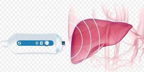
1. Overview of liver hemangioma
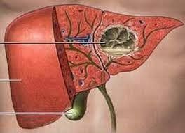
Liver hemangioma is a benign tumor. Hemangiomas can form anywhere in the body, however, the liver is the most common site where hemangiomas appear. Liver hemangioma is a tumor with many blood vessels. This is a rare disease and occurs more often in women than in men. Liver hemangiomas are often discovered incidentally through routine physical examination or other disease.Up to now, the cause of liver hemangioma is still unknown. Many studies suggest that female sex hormones can contribute to hemangiomas in the liver in women.
In the liver, hemangiomas usually appear in the right liver and located in the subcapsular region, with a size of a few mm to 3-4 cm. Patients are usually found to have a single tumor of small size, but there are also cases of patients with multiple hemangiomas or large tumors.
Most patients with liver hemangiomas have no specific symptoms until they are accidentally discovered, especially when the hemangioma is larger than 4cm, the patient may have symptoms such as: pain on the right side of the abdomen. , nausea and vomiting, indigestion, flatulence, abdominal distension, loss of appetite.
Bệnh u máu gan là bệnh lành tính không gây nhiều nguy hiểm cho người bệnh
2. Diagnosis of liver hemangioma
When a patient with liver hemangioma shows symptoms, the doctor can assign the patient to perform the following methods to diagnose the disease:Liver ultrasound: Ultrasound is an imaging technique that uses waves High frequency ultrasound for liver reconstruction. CT scan: CT scan is a computerized tomography technique that uses X-rays to record, process, and build images of liver slices. MRI scan: An MRI scan, also known as magnetic resonance imaging, is an imaging technique that uses radio waves and a magnetic field to recreate images of the liver. Radiographs: Liver hemangiomas can also be diagnosed by radiography. This is a technique to reconstruct the liver image using radioactive isotopes. In addition, to diagnose liver hemangiomas, doctors also appoint patients to perform other methods such as blood tests, liver biopsy. Depending on the condition, the doctor will prescribe the appropriate method.

Chụp MRI giúp đánh giá tình trạng u máu gan
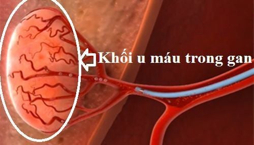
3. Liver hemangioma on ultrasound
Among the diagnostic methods for liver hemangiomas mentioned above, liver ultrasound helps to detect and classify hemangiomas as typical or atypical.Typical liver hemangioma: This is a common form, with the average tumor size being less than 3cm. The image of liver hemangioma on ultrasound is a homonymous and hyperechoic mass, clearly demarcated, and the posterior region is hyperechoic. In the central region of the lesion may be hypoechoic or thin. Atypical hepatic hemangiomas: On ultrasound, atypical liver hemangiomas may be hypoechoic, homophonic or non-echoic. Hypoechoic occurs in the case of fatty liver, hematopoietic tumors can reduce the sound compared to the adjacent parenchyma. Non-homonymous occurs in the case of cirrhosis, liver thrombosis, tumor bleeding, or liquefied tumor, pseudocyst, at this time the image of liver hemangioma on ultrasound is a large mass, non-homonymous, regional hyperechoic periphery, hypoechoic or central hollow. Homophones are rare.
Siêu âm gan là một phương pháp chẩn đoán u máu gan
4. What to do when detecting liver hemangiomas on ultrasound?
Patients should not be too worried when detecting liver hemangiomas on ultrasound, because this is a benign tumor and can be treated. As mentioned above, most cases of hemangiomas in the liver are small in size and do not require treatment.Currently, there are no drugs that can reduce or completely eliminate the tumor. For rare cases of large hemangiomas, in order to prevent the tumor from bursting and bleeding or causing pain, the patient is now treated with one of the methods such as surgical removal of part of the liver hemangioma. or radiotherapy to embolize the artery feeding the tumor, ...
The liver hemangioma on ultrasound can be a very large hypoechoic or non-homogenous mass, depending on the typical or atypical form. Most cases of liver hemangiomas are small in size and do not require treatment. Only treat in case the tumor is large and painful, preventing tumor rupture and life-threatening visceral bleeding.
Although, liver hemangioma is a benign tumor, but when there is a history of liver diseases, the patient should have regular health check-ups to promptly detect the disease, providing a high chance of treatment.

Người bệnh có thể được phẫu thuật cắt bỏ một phần khối u máu gan
When registering for the Liver Cancer Screening and Early Detection Package, the patient will be directly examined by a specialist doctor, will be given tests to assess liver function, screen for hepatitis B, hepatitis C and hepatitis C. diagnostic imaging for malignant tumors. With a system of modern machinery and a team of highly qualified and experienced doctors, Vinmec is committed to the best protection for the health of patients.
Please dial HOTLINE for more information or register for an appointment HERE. Download MyVinmec app to make appointments faster and to manage your bookings easily.