This is an automatically translated article.
Hematoma in the liver is usually the result of trauma, contusion or puncture. The patient presents with abdominal pain, sometimes radiating to the shoulder. Diagnosis of liver injury is made by CT scan or ultrasound. The main method of management is observation, sometimes surgery.
1. Causes of hematoma in the liver
Liver hematomas can be caused by strong impacts (eg, car accidents) or penetrating injuries (eg, stab wounds, gunshot wounds). Liver injury includes subcapsular hematomas, small capsular tears, sometimes deep parenchymal tears, and vascular pedicle rupture.
Liver injuries are classified according to 6 degrees of severity. The immediate consequence of liver injury is usually bleeding. The amount of bleeding may be less or more depending on the nature and extent of the injury. For minor tears, especially in children, the hematoma in the liver may stop on its own. Large lesions in the right abdomen can lead to severe hemorrhage, causing hemorrhagic shock. Mortality rate increases if the patient has severe liver damage.
2. Signs of liver hematoma
Patients with hepatic hematoma present with severe abdominal bleeding, including hemorrhagic shock, abdominal cramps, peritoneal tenderness, and abdominal distention. These symptoms are almost clinically detectable. Accordingly, signs of small hemorrhage or hematoma in the liver lead to abdominal pain and abdominal wall reaction in the right upper quadrant abdomen.
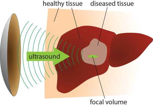
Depending on the severity of the liver injury, the doctor will have an appropriate treatment. Most cases of suspected hepatic hematoma require bed immobilization, intensive resuscitation, and close follow-up with modern diagnostic tools, including computed tomography, ultrasonography. , angiography. If patients with severe hepatic hematoma present with traumatic shock and hypovolemia (pale skin and mucous membranes, fainting or dizziness, difficult or undetectable pulse due to small tachycardia), intervention is recommended. early surgery.

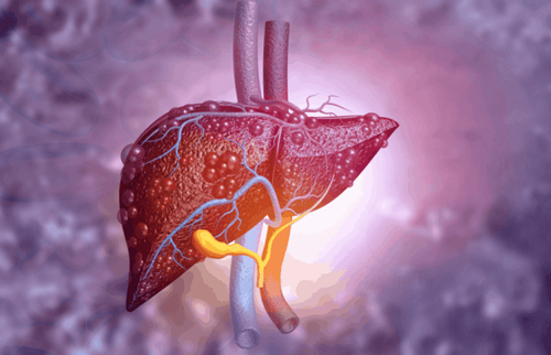
Người bệnh có thể gặp tình trạng đau quặn bụng do tụ máu ở gan
3. Treatment of hematoma in the liver
Patients with liver injury leading to hematoma are often monitored for hemodynamic indicators, vital signs and intervention when there are signs of abnormality during the follow-up period. Severe hepatic hematoma sometimes requires embolization or surgery.
If the patient is hemodynamically stable and there is no indication for laparotomy (eg, perforation of a hollow viscera), vital signs and HCT (Hematocrit) can be monitored. Intervention is needed in patients who are actively bleeding (eg, those with hypotension and shock, who require continuous blood transfusions, or whose HCTs are decreased). For patients with hepatic hematoma with stable vital signs but requiring continuous blood transfusion, angiography and selective embolization should be performed. If the patient's general condition is unstable, laparotomy should be considered.
The success rate in nonoperative treatment of hematoma in the liver for each degree of injury is:
Grade 1 and 2 lesions: Approximately 92% Grade 3: Approximately 80% Grade 4: Approximately 72 % Grade 5: Approximately 62% During surgery, small tears are usually sutured or hemostasis is performed with substances such as oxidized cellulose, fibrin glue, thrombin mixture and gelatin powder. Laparotomy for deeper and more complex lesions is often difficult.
4. Complications due to hematoma in the liver and treatment direction
The overall complication rate for liver hematomas is <7%, but can be as high as 15-20% for severe lesions. Deep parenchymal tears have the potential to lead to bile leaks or the formation of bile ducts. When a bile leak occurs, bile leaks freely into the abdominal cavity or chest. Whereas, cholecystitis is the formation of a bile-filled mass resembling an abscess. Gallstones are usually managed with percutaneous drainage. For biliary fistula, biliary pressure should be reduced through endoscopic retrograde cholangiopancreatography (ERCP), which has a high success rate.
Nội soi mật tụy ngược dòng được sử dụng giảm áp đường mật trong tụ máu ở gan
Abscess occurs in about 3-5% of liver injuries, this condition is usually due to tissue breakdown upon contact with bile. When this complication is suspected in patients with pain, fever, and leukocytosis during the days of hepatic hematoma, a CT scan should be performed to confirm the diagnosis. Abscesses are usually treated by percutaneous drainage; if unsuccessful, laparotomy should be considered.
After treatment, the patient should follow the doctor's instructions regarding the length of stay in the ICU, hospital stay, recovery with diet, rest, or limited activity after discharge.
Currently, many new methods in the treatment of injuries and hematomas in the liver have been applied, such as laparoscopic surgery, causing embolism when treating bleeding,... When the symptoms of hematoma in the liver are noticed, , suspected due to injury, the patient needs to see a specialist for timely treatment and treatment.
Please dial HOTLINE for more information or register for an appointment HERE. Download MyVinmec app to make appointments faster and to manage your bookings easily.