This is an automatically translated article.
The article was professionally consulted by Specialist Doctor I Vo Cong Hien - Radiologist - Radiology Department - Vinmec Nha Trang International General Hospital. The doctor has many years of experience in the field of diagnostic imaging.Epiploic appendagitis is a rare inflammatory condition that affects the small fat-filled sacs (fatty manes) on the colon. Although people with end-stage steatorrhea can experience severe lower abdominal pain, nausea, and vomiting, doctors can easily treat it with pain relief and antibiotics.. Today, thanks to the use of ultrasonography and CT scan in the evaluation of acute abdominal pain, steatorrhea can be detected by characteristic imaging features.
1. What is steatorrhea?
Fatty colonitis is an inflammation of the small pouches in the colon. These vesicles are called epigenetic appendages or fat manes. They are part of the omentum of the colon, which is supplied with blood from one or two arterioles and one vein.These fatty manes help the body absorb nutrients, protect the blood vessels in the colon. Most people have about 50-100 fat manes, 0.5-5cm long, they are located on the outer surface and parallel to the longitudinal muscular band of the colon. Fatty mane of the colon occurs if the blood supply to these fatty deposits is cut off. The lack of blood flow causes fatty tissues to become inflamed, leading to severe lower abdominal pain.
Colitis fatty mane can be confused with other diseases such as:
Appendicitis . Diverticulitis. Mesenteric lymphadenitis. However, unlike the above conditions, steatorrhea is a benign disease that usually does not require surgical treatment. Today, ultrasound and CT scan play an important role in the diagnosis of this condition.
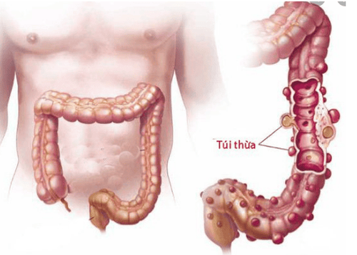
Viêm bờm mỡ đại tràng là tình trạng viêm các túi nhỏ trên đại tràng.
2. What causes colonic fatty mane?
There are two types of steatorrhea: primary steatorrhea and secondary steatorrhea. Both types are associated with loss of blood flow to fatty manes, but they have different causes.2.1. Primary steatitis of the colon
Primary steatorrhea occurs when the blood supply to the fatty deposits is cut off. Sometimes, fatty manes become twisted, compressing blood vessels and preventing blood flow. In some cases, blood vessels can suddenly collapse or have a blood clot that blocks blood flow to the fatty mane.2.2. Secondary steatorrhea
The etiology of secondary steatitis may be diverticulitis, appendicitis, cholecystitis, etc. The colonic fatty mane is seen on CT scan only when it is inflamed or surrounded by fluid. .3. Who often suffers from colonic fatty mane?
It is more common in men between the ages of 40 and 50. Other possible risk factors include:Obesity : May increase the number of fatty manes. Overeating: May alter blood flow to the intestinal tract. Less exercise.

Viêm bờm mỡ đại tràng thường phổ biến hơn ở nam giới trong độ tuổi 40 và 50.
4. Symptoms of Fatty Colitis
Some possible symptoms of steatorrhea fasciitis:The patient feels pain in the lower abdomen, usually more pain on the left side than on the right, pain increases with movement.
Fever. Nausea, vomiting. Diarrhea . Constipation. Anorexia.
5. Diagnosis of colonic fatty mane
Diagnosis of steatorrhea usually involves excluding other conditions with similar symptoms, such as diverticulitis or appendicitis.Therefore, if the doctor suspects that the patient has steatorrhea, a blood test may be performed to see the white blood cell count. Patients with steatorrhea often have normal or slightly elevated white blood cell counts.
If the white blood cell count is significantly elevated, it is most likely a sign of other inflammatory conditions such as diverticulitis or appendicitis.
The doctor may also use diagnostic imaging tests, such as a CT scan or ultrasound, to examine the organs inside the abdomen. The normal fatty mane is not visible on the CT scan.
6. Image of colonic fatty mane on CT scan
CT scan is the most effective diagnostic modality for steatorrhea. Here are some common pictures of fatty mane on CT scan:Appears a dense image of oval fat mane, size 2 - 4cm, adjacent to colon surrounded by these inflammatory lesions. Occasionally there is thickening of the adjacent parietal peritoneum due to widespread inflammation. A central contrast-enhanced nodule may be present due to venous embolism. The inflamed fatty mane is usually located on the anterior surface of the sigmoid or descending colon, but it can be anywhere along the circumference of the colon.

CT scan là phương pháp chẩn đoán hiệu quả nhất đối với viêm bờm mỡ đại tràng.
7. Methods of treatment of colonic fatty mane
Fatty colonitis is generally considered a benign disease. This means it should go away on its own without treatment. In the meantime, your doctor may recommend an over-the-counter pain reliever, such as acetaminophen (Tylenol) or ibuprofen (Advil). In some cases, patients may need to take antibiotics. The patient's symptoms should begin to improve within a week of taking the drug.Surgery may be considered in the event of complications or recurrences.
There is no specific diet that people with end-stage steatorrhea should or should not follow. Obesity and overeating can be risk factors, so developing a balanced, portion-controlled diet to maintain a healthy weight can help prevent pain.
Cases of secondary steatorrhea usually resolve after the underlying cause is treated. Depending on the condition, the patient may need to have the appendix or gallbladder removed, or other intestinal surgery.
Please dial HOTLINE for more information or register for an appointment HERE. Download MyVinmec app to make appointments faster and to manage your bookings easily.
Reference sources: healthline.com, Sinhvienykhoa115.wordpress.com, radiopaedia.org


