This is an automatically translated article.
The article is professionally consulted by Master, Doctor Nguyen Hong Hai - Doctor of Radiology - Department of Diagnostic Imaging and Nuclear Medicine - Vinmec Times City International Hospital
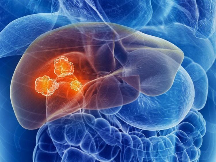
Liver abscess is a condition where the liver cells are destroyed, forming a pus pocket. With the advancement of science and technology today, diagnosing liver abscess becomes easier. In particular, abdominal and pelvic scans are often indicated for hepatobiliary diseases, including liver abscess.
1. Bacterial liver abscess
1.1. Pathogens Bacteria invade the liver, causing inflammation and necrosis of liver cells, creating pus-filled foci. Commonly found are Staphylocoque bacteria, Streptocoque bacteria, intestinal bacilli Salmonella, even anaerobic bacteria and hair fungus (Leptothrix).
They enter the liver by many different routes as follows:
By biliary tract: This is a complication of biliary tract diseases, for example gallstones, worms in the bile ducts, diseases that cause long-term stenosis and cholestasis. : Bacteria from all intra-abdominal infections - especially abscesses, can travel through the veins to the liver By lymphatic routes, hepatic arteries: Bacteria from any site of infection in Any body can cause liver abscess. Patients may find scattered pus in the lungs, brain, muscles, kidneys, and in the liver. 1.2. Diagnosis and treatment The characteristic of bacterial liver abscess is that there are many pus-filled foci of different sizes, scattered in the liver. Sometimes only one lobe is seen, but there are still many large and small clusters concentrated in one block.

Diagnosis of bacterial liver abscess is mainly based on the clinical presentation of the disease. Symptoms such as right lower quadrant pain, enlarged liver, painful intercostal pressure, X-ray or ultrasound with pus in the liver... are warning signs of aggravation.
The principle of treatment is to use strong antibiotics in combination with surgical intervention depending on the nature of the cause of the liver abscess.
2. Amoeba liver abscess
2.1. Characteristics of amoebic liver abscess caused by bacillus dysentery penetrate the intestinal wall, follow the venous capillaries and then carry the veins to the liver, destroy liver cells and create large pustules. Features of this type of liver abscess are:
Only amoeba is the causative agent, there are no bacteria in the pus, if any, it is due to superinfection. Therefore, some experts also call this as a sterile liver abscess. Usually only a large pus-filled fovea, 2-3 foci of pus are rarely seen, mainly in the right lobe because the burden vein on this side is larger, straighter and shorter than the pus. , do not rot and often have a distinctive chocolate color. When an infection occurs, the color will change. 2.2. Symptoms Clinical manifestations of liver abscess caused by amoeba include:
High fever with chills Hepatomegaly, pain Painful intercostal pressure

Subclinical:
X-ray film shows that the right diaphragm is high, has little movement, the right flank angle is blurred. Blood tests show high leukocytes and erythrocyte sedimentation rate... Note, there are cases of the disease. The abscess is very large, but the symptoms are poor.
2.3. Diagnosis and treatment definite diagnosis Besides clinical symptoms, the doctor can apply methods such as X-ray, hepatic angiography or umbilical vein, laparoscopy, liver scintigraphy, puncture , abdominal scan and liver ultrasound. Currently, thanks to ultrasound, other techniques are rarely used. However, there are also cases where ultrasonography can completely detect amoeba, as well as not definitively identify liver abscess.
Differential diagnosis Amoebic liver abscess in the non-purulent stage needs to be differentiated from many other diseases, such as liver cancer, perforation of the stomach and duodenum, acute pancreatitis, perirenal abscess and some diseases. other
Treatment Currently, there are combined measures to treat amoeba-induced liver abscess, including specific drugs, removing pus-filled foci once formed, and combining antibiotics.
3. Abdominal and pelvic scan to diagnose liver abscess
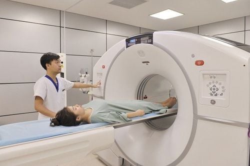
Computerized tomography (CT scan) is more and more widely used and indicated in Vietnam. This is a very valuable technical means, indispensable in clinical practice. However, the analysis and evaluation of abdominal and pelvic scans is relatively difficult and complicated.
Abdominal and pelvic scans are often indicated in the diagnosis of diseases of the abdomen and pelvis. The majority were pathologies of the peritoneum, retroperitoneal space and related viscera, vascular disease, bone and soft tissue disease of the abdominal - pelvic wall. In particular, abdominal and pelvic scans are also performed for liver problems, such as benign/malignant tumors, liver infection or injury.

Examine the liver with contrast-enhanced CT to increase contrast, distinguish between the density and lesions, especially of liver tumors. The effectiveness of contrast-enhanced abdominal and pelvic scans depends on many factors, such as contrast concentration and speed, body weight, time of procedure, etc. The current popular technique is the study of increased kinetics. X-ray at different stages, helps to increase the sensitivity in diagnosis and clearly distinguish different types of liver tumors.
On CT film, liver abscess caused by pyogenic bacteria shows a mass occupying the place with reduced density, round shape, irregular border, and wall becomes radiolucent after contrast injection. On contrast, a double cover is sometimes seen due to a central hypoattenuating area. There may be one or more abscesses (collected, clustered), segmental margins. Typically air bubbles or fluid levels. Distinguishing a liver abscess from a cyst or a necrotic tumor by scanning the abdomen and pelvis is sometimes difficult.
Computed tomography image of amoebic liver abscess is nonspecific, often showing a circular or oval lesion. Observe the non-contrast film, the lesion is hypoattenuated. With contrast film, the lesion is enhanced border, no central increase, irregular border, can form the inner wall. Extrahepatic manifestations include pleural effusion, perihepatic fluid, and gastric, colonic, and retroperitoneal changes.
There are also liver abscesses caused by large flukes (common in the central part of our country), often involving the right lobe, with non-specific symptoms. Abdominal and pelvic scan showed many small lesions, about 2-3 cm in size, clustered and clustered. Observation of non-contrast film showed hypoattenuating lesions. For films with contrast, it will show a borderline enhanced enhancement, with an inner wall.
Liver infection can be life-threatening if not diagnosed and treated promptly. Therefore, screening for liver infection on computed tomography is very necessary.
Currently, computed tomography is an imaging method performed routinely at Vinmec International General Hospital. The computerized tomography procedure at Vinmec is carried out methodically under the guidance of the medical team of the Department of Diagnostic Imaging. In addition, Vinmec is now equipped with the most modern CT scanner, meeting international standards for true and clear images, helping doctors to accurately diagnose the disease and the stage of the disease, thereby effective treatment, creating a sense of safety for the patient.
Customers who need to be examined and treated at Vinmec International General Hospital, please register for an online examination on the Website for the best service.
Please dial HOTLINE for more information or register for an appointment HERE. Download MyVinmec app to make appointments faster and to manage your bookings easily.


