This is an automatically translated article.
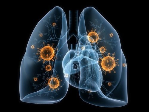
The lungs are the central organs of the respiratory system. Understanding the anatomy of the lungs and pleura helps patients better understand this organ and take effective measures to protect the lungs against pathogens.
1. What is the function of the lungs?
Lungs are organs of the respiratory system, where air exchange between the body and the environment. The lungs are enclosed in a serosa consisting of 2 leaves. Lung properties are elastic, spongy and soft. Each person has 2 lungs, made up of lobes. The right lung is usually larger than the left lung. The capacity of each lung is about 5000ml during forced inhalation.
The function of the lungs is to exchange air and blood. This process is carried out on the entire inner surface of the bronchi. To remove foreign objects, the lining covering the alveoli is always vibrating. Lung cells play a role in maintaining epithelial and endothelial cell survival. Lung cells form a barrier that prevents molecules and water from entering the interstitium. At the same time, lung cells are also involved in the transport and synthesis of many substances in the body.
2. Embryology
Before learning about lung anatomy, everyone should understand the formation of the lungs from the embryonic stage:
The germ of the respiratory organs is a long groove, appearing in the 4th week of the embryo. The germ tube is covered by endodermal tissue (this organization later develops into the epithelial layer of the respiratory organs). The first part of the germ tube develops into the larynx and trachea. From the tail of the germ tube, on the 2 side walls grow 2 buds of the right and left lungs. Initially, the 2 lung buds develop symmetrically and symmetrically, but from 6 to 7 weeks, the lungs begin to divide into lobes. The right lung is divided into 3 lobes, the left lung is divided into 2 lobes.
From the 6th week of pregnancy, the development of the components of the bronchi complete. An airway consisting of terminal bronchi has been formed. As the lungs develop, they will protrude from the sides into the thoracic cavity to later form the pleural cavity.
By the 6th week of pregnancy, the left 6th aortic arch will form the left and right pulmonary arteries. Each pulmonary artery is closely related to the main bronchus, which develop into branches accompanying the corresponding bronchus. These pairs of arteries and bronchi enter the lobes of the lung, branch off into the segments, and develop into the lung parenchyma. As the lungs develop, pairs of arteries and bronchi are centrally located each, subdividing into branches.
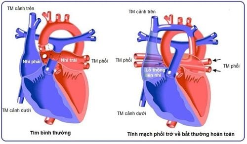
The major pulmonary veins are developed from the atrial germ. The veins in the lungs are made up of the mesenchymal tissue layer. The pulmonary veins receive pleural branches and a network of blood vessels that develop from the top of the bronchial tree. When going to the periphery, the veins in the lungs form the interlobar veins, which run in the connective tissue layer between the lobes of the lung. The veins in the lobes of the lung merge, forming the lobe veins. The segmental veins merge to form the great vein, the pulmonary vein near the hilum. These veins continue to merge, forming the superior and inferior pulmonary veins.
Watch now: Overview of common lung diseases

Sự hình thành của phổi được bắt đầu từ giai đoạn phôi thai
3. Lung Anatomy
In cardiopulmonary anatomy, the lung consists of an external and an internal form.
3.1 Extracorporeal appearance The lung has a semi-conical shape, suspended in the pleural space by the pulmonary ligaments and the bronchi. Lungs have 3 sides, 1 apex, and 2 edges. The outer surface is convex, pressed against the chest wall; the mediastinum is the bilateral limit of the mediastinum; The lower surface (bottom of the lung) is pressed against the diaphragm.
The external structure of the lung includes:
Bottom of the lung: Located close to the diaphragm, through the diaphragm, related to the abdominal organs, especially the liver; Lung apex: Protrudes from rib 1. Posteriorly, apex of lung is level with posterior end of rib 1. Anteriorly, apex of lung is about 3cm above medial clavicle; The ribs: The common point of the 2 lungs is that they are close to the inside of the chest, with the impressions of the ribs. The characteristics of each lung are: Right lung has 3 lobes (upper, middle and lower lobes), left lung has 2 lobes (upper and lower lobes); Inner surface: Slightly concave, consisting of 2 parts: the vertebral column (related to the spine) and the mediastinal part (contains the viscera in the mediastinum). Between the inner surfaces of the two lungs is the hilum. Behind the hilum, there is a single venous fissure and compression of the esophagus in the right lung, the aortic groove in the left lung. Above the hilum, there is the brachiocephalic sulcus and the subclavian artery; Borders: Consists of anterior margin (formed by the flank and diaphragm) and lower border (consisting of two segments, curved and straight). 3.2 Inner shape In lung anatomy, the lung is composed of components that pass through the hilum, gradually dividing in the lung. These are: the bronchial tree, the pulmonary artery, the pulmonary vein, the bronchial artery, the bronchial vein, the lymphatics, the connective tissues, and the nerve fibers.
Characteristics of the internal shape of the lung are as follows:
Bronchi tree: The main bronchus enters the hilum and divides into lobar bronchi. Each lobar bronchus carries air for 1 lobe of the lung, further dividing into segmental bronchi to carry air for 1 lung segment. The segmental bronchus divides into the subsegmental bronchus, dividing several times to the lobar bronchi - conducting air for 1 lobe of the lung; Pulmonary artery: The trunk of the pulmonary artery passes from the foramen of the pulmonary artery of the right ventricle to the posterior border of the aortic arch, dividing into the left pulmonary artery and the right pulmonary artery. The left pulmonary artery crosses in front of the left main bronchus, entering the hilum above the left upper lobe bronchus. The right pulmonary artery enters the right hilum in front of the main bronchus, exits, and goes behind the bronchus; Pulmonary vein: The alveolar reticulum drains into the perilobular vein, continues to form progressively larger trunks, to the intersegmental or intrasegmental veins, the lobar vein, which merges into 2 veins On each side of the lung, it carries oxygen-rich blood back to the left atrium. The pulmonary venous system has no valves; Bronchial arteries and veins: Supply nutrients to the lungs. The bronchial artery is the lateral branch of the aorta. The bronchial veins empty into the single and pulmonary veins; Lymphatic: Consists of many lymphatic vessels running in the lung parenchyma, then emptying into the pulmonary lymph nodes, emptying into the upper and lower tracheal nodes at the bifurcation of the trachea; Nervous system: Nerves to the lungs include the sympathetic nervous system and the parasympathetic nervous system.
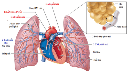
Hình thể trong của giải phẫu phổi
4. Anatomy of the pleura
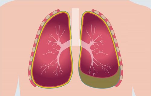
Besides lung anatomy, pleural surgery is also a matter of great interest to many people. The pleura is a vascular bar consisting of 2 leaves: the visceral pleura and the parietal pleura. Between the 2 leaves is the pleural cavity. Specific features are as follows:
Visceral pleura: Covers the entire surface, adheres tightly to the lung parenchyma, splenic into the interlobular slits. In the hilum, the visceral pleura fan out, adjacent to the parietal pleura; Parietal pleura: Lines the inside of the chest cavity, forming the pleural sac. Parietal pleura includes: mediastinal pleura, costal pleura, and diaphragmatic pleura. The apex of the pleura is the parietal pleura that corresponds to the apex of the lung. The pleural space is made up of 2 parts of the parietal pleura, including the diaphragmatic space and the mediastinal space; Pleural cavity: Is a virtual space, located between the visceral pleura and parietal pleura. Each lung has a closed, separate and non-connecting pleural cavity. Blood vessels and nerves of the pleura:
Arteries: The parietal pleura is supplied with blood by branches of adjacent arteries (intercostal arteries, internal thoracic arteries, mediastinal branches, arteries of the diaphragm) . The visceral pleura is supplied with blood from the bronchial arteries; Veins: Along with arteries; Nerves: The costal pleura is innervated by the intercostal nerves; the costal and mediastinal pleura are innervated by sensory branches of the phrenic nerve; The visceral pleura is innervated by the pulmonary plexus. Lung and pleural anatomy information helps readers better understand the structure of the parts of this respiratory organ. Everyone should pay attention to stay away from tobacco smoke, polluted environment, eat healthy foods and exercise,... to prevent lung diseases.
Follow Vinmec International General Hospital website to get more health, nutrition and beauty information to protect the health of yourself and your loved ones in your family.
Please dial HOTLINE for more information or register for an appointment HERE. Download MyVinmec app to make appointments faster and to manage your bookings easily.


