This is an automatically translated article.
Posted by Doctor Tran Truong Giang - Intensive Care Unit - Vinmec Times City International HospitalEmergency echocardiography at the patient's bedside is a necessary technique to help doctors quickly diagnose some cardiovascular disorders and thereby provide more effective treatment.
1. Outline
Bedside emergency echocardiography is an essential non-invasive exploratory technique in the emergency resuscitation of critically ill patients, helping clinicians quickly diagnose a number of cardiovascular disorders, thereby provide more effective and effective treatment.2. Indication of emergency bedside echocardiography
Myocardial infarction Pericardial effusion, hemopericardial effusion Heart valve disease Shock condition: cardiogenic shock, septic shock ... Monitor the effectiveness of treatment methods3. Contraindications to bedside emergency echocardiography
There are no contraindications.4. Prepare an emergency echocardiogram at the bedside
Performer: 1 doctor and 2 nurses Doctor trained in echocardiography, vascular: Wear hat, mask, wash hands. Sit to the right of the patient. The right hand holds the transducer, the left hand adjusts the buttons of the ultrasound machine. Nursing: wear a hat, wear a mask. 01 The nurse monitors vital functions, ensures breathing and intravenous lines for the patient during the ultrasound. 01 Nursing assistant to the doctor during the procedure: Change the patient's position. Means Ultrasound machine with vascular ultrasound function. Monitor monitors vital functions: heart rate, SpO2, breathing rate, blood pressure. Ultrasound gel: 1 bottle Clean sterile gauze: 1 pack Examination gloves: 3 pairs Surgical caps: 3 pieces Surgical masks: 3 pieces Quick hand sanitizer Electrocardiogram monitoring electrodes when doing ultrasound: 3 pieces machine maintenance cost Machine depreciation expense Patient Explain to patient and patient's family the benefits of bedside vascular ultrasound. The patient lies on his back, depending on the position of the ultrasound, there are different positions. Simultaneous electrocardiogram during ultrasound. Patients with mechanical ventilation must pay attention to ensure the patient's respiratory status during the ultrasound. Patients with infusion of vasopressors must pay attention to ensure the intravenous line during the ultrasound. Medical record Record the order of vascular ultrasound. Record the measured parameters on the ultrasound results sheet and paste it on the medical record.5. Emergency echocardiography procedure
Check the records: Check the indications, contraindications and the written consent to participate in the techniqueCheck the patient again: The vital functions can be performed.
Technical implementation:
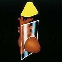
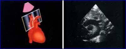
5.1 2D ultrasound Allows to examine the structure of the working heart a). Cross section of the left sternum. The transducer is placed at the left sternal border, 3rd or 4th, or 5th intercostal space.
Longitudinal view
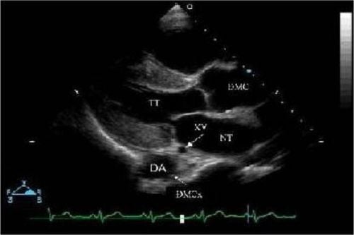
Mặt cắt cạnh ức trái trục dọc
Can detect pathological manifestations of the heart:
Heart valve prolapse: Aortic valve, HL valve HL valve stenosis, thickening, calcification of HL valve.. Thrombosis in left atrial chamber, left atrial mucinous tumor... Abnormal movement of VLT, posterior wall of left ventricle in myocardial infarction, pericardial effusion causing acute tamponade. HL valvular ligament rupture Pericardial effusion: There is an echocardiographic gap in the posterior wall of the left ventricle, if the number is large, the ultrasound space can be seen in the anterior wall of the right ventricle. Horizontal cross-section:
At right angles to the longitudinal axis of the heart (rotate the transducer 900 clockwise), there are 3 cross-sections from top to bottom (section through the origin of great vessels, cross-section through the HL valve). , cross-section of the muscle column)
Cross-section through the base of the great blood vessels: The aorta is observed, the aortic valve is shaped like a ―Y―, the left atrium, the right atrium, the interventricular septum, the tricuspid valve, the right ventricular ejection chamber , pulmonary valve, pulmonary artery stem, 2 branches of right pulmonary artery and left pulmonary artery, left coronary artery.
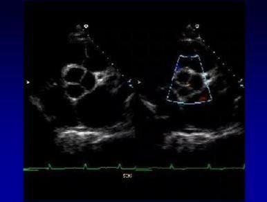
Mặt cắt cạnh ức trái trục ngang qua gốc mạch máu lớn
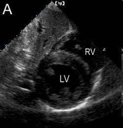
Cross section of the muscle column: Seeing 2 muscle columns (anterior lateral and posterior middle), the right ventricle is smaller than the left ventricle and located anterior to the left ventricle, with The descending (transverse) aorta can be seen behind the left ventricle.
Mô hình mặt cắt cạnh ức trái trục ngang thấp dưới van hai lá
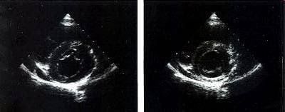
Mặt cắt cạnh ức trái trục ngang thấp dưới van hai lá
Aortic valve: Thickening of each aortic valve can be seen, the aortic valve has only 2 leaflets. Valve HL: Foot valve, valve stenosis HL Abnormal movement of the VLT, left ventricular posterior wall in myocardial infarction, pericardial effusion causing acute cardiac tamponade. Pericardial effusion: there is an echocardiographic void in the posterior wall of the left ventricle, if the number is large, the echocardiogram can be seen in the anterior wall of the right ventricle. Pulmonary stenosis. Views from the apex of the heart:
The patient lies supine, the transducer is placed at the apex towards the base of the heart.
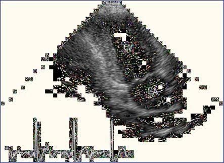
Four-chamber view:
Surveying the longitudinal structure of the heart (2 ventricular chambers, interventricular septum (VLT), two atrial chambers, HL valve, BL valve, pulmonary veins emptying into the NT.

Mặt cắt 4 buồng từ mỏm tim
From the 4-chamber view, rotate the probe 900 clockwise to get a 2-chamber view, surveying the left ventricle, left atrium, left ventricular anterior wall, left lower wall.
Mặt cắt 2 buồng từ mỏm tim
Heart valve prolapse: HL valve, BL valve HL valve stenosis, thickening, calcification of HL valve.. Thrombosis in left atrial chamber, left atrial mucinous tumor... Abnormal movement of VLT, left ventricular wall in myocardial infarction, pericardial effusion causing acute tamponade. HL valvular ligament rupture Pericardial effusion: If the number is high, the ultrasound space can be seen in the right and left ventricle walls (4-chamber view), the echocardiographic space in the lower left ventricle (2-chamber view). ). Subcostal view:
Patient lying supine, knees slightly flexed, transducer placed in epigastrium below the sternum
4-chamber view:
See cardiac structures similar to 4-chamber view from the apex

Mặt cắt 4 buồng dưới bờ sườn
Can cut along the heart bottom to see the aorta and pulmonary artery body, cross the HL valve, across the muscle column, across the inferior aorta and the right atrium.
Mặt cắt dưới sườn trục ngang, ngang qua cột cơ
Heart valve prolapse: HL valve, BL valve HL valve stenosis, thickening, calcification of HL valve.. Thrombosis in left atrial chamber, left atrial mucinous tumor... Abnormal movement of VLT, left ventricular wall in myocardial infarction, pericardial effusion causing acute tamponade. Rupture of the HL valve ligament Pericardial effusion: If the number is high, the ultrasound space can be seen in the right and left ventricular walls (4-chamber view). Periventricular left ventricular (horizontal view). Transsternal cross-sections of the supraclavicular
In longitudinal axis: The aortic arch and trunk branches of the brachial artery, left carotid artery, left subclavian artery, ascending aorta, descending aorta, isthmus, and right pulmonary artery can be seen.
Mặt cắt trên hõm ức theo trục dọc

Mặt cắt trên hõm ức theo trục ngang
Coarctation of the aorta Aneurysm, dissection of the aorta in the loop. 5.2 Transvaginal ultrasound Ultrasound waves are perpendicular to cardiac structures, helping to measure the thickness and width of these structures. The transducer is placed on the left border of the sternum in the 3rd or 4th intercostal space, the transducer makes an angle of 800 - 900 with the thoracic plane.
Location of the ventricular cross-section:
Left parasternal map (long or short axis) is the most standard position to measure the size of the left ventricle on echocardiography (according to the American Society of Echocardiography).
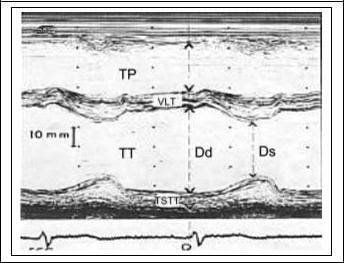
Siêu âm TM ngang thất trái
At the end of diastole (beginning of Q wave of the QRS complex on ECG) right ventricular diameter, interventricular septal wall thickness, left ventricular diameter, left ventricular posterior wall thickness
At the end the systolic period (measured where the ventricular septum reaches its maximum thickness) septal thickness, left ventricular diameter, left ventricular posterior wall thickness.
Left ventricular ejection volume, from which cardiac output can be calculated.
Can detect pathological manifestations of the heart:
Abnormal mobility of the HL valve: parallel movement in HL
VLT thickness (bt: diastolic 7.5 ± 1mm; systolic 10 ± 2 mm )
Left ventricular posterior wall (bt: diastolic 7.0 ± 1 mm; systolic 12 ± 1 mm)
Abnormal movement of VLT, posterior wall of left ventricle: decreased mobility, paradoxical mobility in myocardial infarction , pericardial effusion causing acute cardiac tamponade.
Right ventricular diameter (bt 16 ± 4mm)
Left ventricular diameter (bt: Diastolic 46 ± 4 mm, systolic 30 ± 3 mm)
Left ventricular diastolic volume (101 ± 17 ml) and systolic ( 37 ± 9 ml), left ventricular ejection volume.
Left ventricular contraction fraction (%D: 34 ± 6)
Left ventricular ejection fraction (EF: 63 ± 7 %)
Pericardial effusion: if the echogenic space is posterior to the left ventricle during systole , diastole.
Cross-sectional position of the aortic valve and left atrium:
From front to back, the structures of the anterior thoracic wall, right ventricular anterior wall, right ventricular ejection chamber, anterior wall of the aorta are connected by the ventricular septum, the connection continuity of the HL-aortic valve, the left atrial chamber, the posterior wall of the left atrium.
Only 2 of the 3 sigmoid leaflets of the aorta can be observed: the right coronary leaflets and the non-coronary leaflets. Mobilize the aortic sigmoid valves when open to form a 'box shape'. This view allows the measurement of:
Aortic end-diastolic diameter (bt: 28 ± 3 mm)
Aortic valve opening amplitude
Left atrial end-systolic diameter (bt: 31 ± 4 mm)
Detectable Cardiac pathological manifestations:
Heart valve prolapse: HL valve, BL valve HL valve stenosis, thickening, calcification of HL valve.. Thrombosis in left atrial chamber, left atrial mucinous tumor... Abnormal movement of heart VLT, left ventricular wall in myocardial infarction, pericardial effusion causing acute tamponade. Pericardial effusion: If the number is large, the ultrasound space can be seen in the right and left ventricle walls (4-chamber view). Periventricular left ventricular (transverse section).

Mặt cắt TM ngang van động mạch chủ và nhĩ trái

Công thức tính tần số sóng siêu âm
θ: angle formed by the incident ultrasound beam fi and the direction of movement of the structure. In cardiology the structure is blood flow, represented by red blood cells
C: velocity of ultrasound in biological tissues (1560 cm/sec). Purpose of doppler ultrasound: non-invasive hemodynamic investigation
Types of doppler ultrasound: Pulsed Doppler, continuous doppler, color doppler (a special type of pulsed doppler). Pulsed Doppler: The emitted and received ultrasound waves are performed by a single crystal, so the ultrasound beam is emitted intermittently so that the transducer picks up echoes after a short time delay. depends on the depth to be probed. Continuous Doppler: The emitted and received ultrasound waves are performed by 2 different crystals of the transducer, so there is no restriction on blood speed. Color Doppler: Is a pulse doppler that the speed and direction of blood flow are shown in different colors with different intensity. By convention, when flow is directed toward the transducer, it is red, and blue when the flow is away from the transducer. Investigate normal flow:
HL flow:
Best recorded in 4-chamber view from the apex Diastole: Two positive waves, including E wave (left ventricular pre-diastolic filling wave) and A wave (filling wave when left atrium contracts)

Doppler xung qua dòng chảy van hai lá
Usually recorded in a 5-chamber view from the apex, or in the right parasternal view, or the suprasternal view shows rapid ascending and descending systolic waves. The spectrum is positive or negative depending on the cross-sectional position. Cardiac manifestations can be detected: aortic regurgitation (regurgitation in diastole)

Doppler xung qua dòng chảy van động mạch chủ
Usually the 4-chamber view from the apex, the left sternal side view, the 4-chamber view below the costal margin. The spectrum of the tricuspid valve line is the same as that of the HL line (positive flow spectrum). During systole, regurgitation can be seen BL (negative flow spectrum).

Doppler xung qua van ba lá
Pulmonary valve flow:
Usually recorded in left parasternal view. During systole, the spectrum has negative currents. Diastole can have a spectrum of pulmonary valve regurgitation (positive current spectrum). Can detect pathological manifestations of the heart: Pulmonary valve regurgitation (regurgitation in the diastolic period) Vinmec International General Hospital with a system of modern medical facilities and equipment along with With a team of experts and doctors with many years of experience in medical examination and treatment, patients can rest assured that they will be examined and treated at the Hospital.
Please dial HOTLINE for more information or register for an appointment HERE. Download MyVinmec app to make appointments faster and to manage your bookings easily.
SEE MOREWhat is special about 4D echocardiography at Vinmec? What is the significance of echocardiography in the examination and detection of cardiovascular diseases? What is a transthoracic echocardiogram?