This is an automatically translated article.
The article is professionally consulted by Master, Doctor Nguyen Minh Son - Interventional Cardiologist - Department of Medical Examination & Internal Medicine - Vinmec Nha Trang International General HospitalHeart valve disease often has no obvious symptoms, most causes are congenital, a few are acquired. Echocardiography plays a very important role in the diagnosis of valvular heart disease. If detected and intervened late, the disease can cause dangerous complications such as myocardial infarction, heart failure.
1. What is echocardiography?
Echocardiography is a method that uses sound waves to create images of the heart. The images obtained through echocardiography will help doctors understand specific conditions of the heart such as the functioning of the heart muscle, heart valves, structural abnormalities of the heart. From there, it is possible to diagnose and identify cardiovascular diseases.
There are two types of echocardiography: transthoracic echocardiography and transesophageal echocardiography. Most of the time, to diagnose common heart diseases, doctors will order a transthoracic ultrasound. For cases that need more specific results on the morphology of the heart valves, the size of the catheters before performing heart surgery, dilation procedures ... the doctor will appoint a transvaginal ultrasound manage. To evaluate coronary artery perfusion function, left ventricular contractility will use stress echocardiography, examining ventricular wall motion during rest and during myocardial stress.

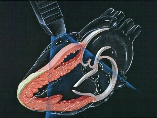
Siêu âm tim để đánh giá chức năng hoạt động của các bộ phận trong cấu tạo của tim
2. Echocardiography to diagnose valvular disease
During echocardiography, doctors will use ultrasound waves to observe the heart structure, assess the heart's functioning, specifically:
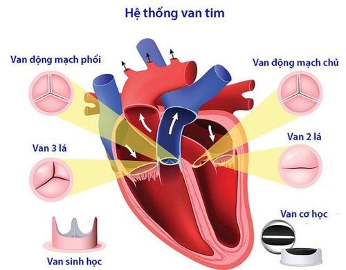
Shape, size of the heart Size, thickness, movement of the heart walls Contraction strength of the heart muscle Valve morphology, movement of the leaflets Presence of tumors Inflammation around the heart valves Problems in the pericardium, large blood vessels , blood clots in the heart chambers... Through observing the morphology of the heart valves, the movement of the leaflets, the doctor will determine whether the patient has heart valve diseases, including stenosis and regurgitation. heart valve.
2.1. Echocardiographic evaluation in valvular stenosis Confirm diagnosis of valvular stenosis Determine severity of valvular stenosis Determine cause and mechanism of valvular stenosis Check left ventricular function Evaluate chamber response to cardiac condition long-term pressure overload Determine pulmonary artery pressure Identify other associated valve injury
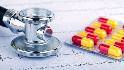
Siêu âm giúp chẩn đoán và đánh giá các bệnh van tim
2.2. Echocardiographic evaluation in valvular regurgitation Definite diagnosis of valvular regurgitation Determining the severity of valvular regurgitation Determining the cause of regurgitation Cardiac chamber size due to volume overload Assess cardiac function left ventricular contraction Determine pulmonary artery pressure Differentiate acute or chronic valvular regurgitation
3. Echocardiography procedure
The patient will lie on the bed, pull up the shirt, the doctor will proceed to attach the electrode patches to the body. These electrode pads will help to measure the electrocardiogram at the same time as the echocardiogram. A special gel will be applied to the patient's body to increase the transmission of sound waves. This special gel also removes air between the skin and the ultrasound transducer. The doctor will move the transducer on the patient's chest, sound waves will help create images of the heart muscle and heart valves. The doctor will observe this image through a display screen. It is a transthoracic echocardiogram.

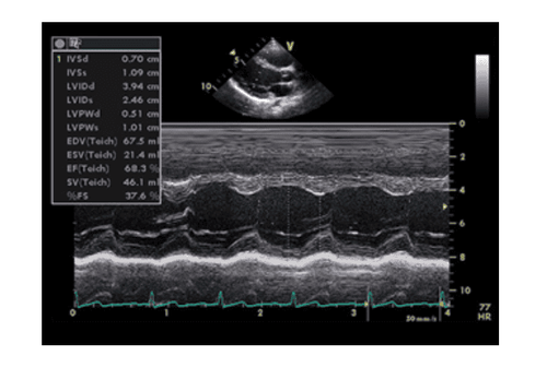
Bệnh nhân tiến hành siêu âm tim theo hướng dẫn của bác sĩ
For transesophageal echocardiography, the patient will be numbed with a gel or spray. The doctor will then insert the probe into the esophagus. Anesthesia will help reduce discomfort. However, the patient needs to fast for a few hours before having a transesophageal echocardiogram.
Through echocardiography, the doctor can diagnose heart valve disease, assess its severity and plan appropriate treatment. Early intervention to treat heart valve disease is very important, minimizing the risk of life-threatening complications.
To protect cardiovascular health in general and detect early signs of myocardial infarction and stroke, customers can sign up for Cardiovascular Screening Package - Basic Cardiovascular Examination of Vinmec International General Hospital . The examination package helps to detect cardiovascular problems at the earliest through tests and modern imaging methods. The package is for all ages, genders and is especially essential for people with risk factors for cardiovascular disease.
Please dial HOTLINE for more information or register for an appointment HERE. Download MyVinmec app to make appointments faster and to manage your bookings easily.