This is an automatically translated article.
The article is professionally consulted by Doctor II Nguyen Quoc Viet - Department of Medical Examination & Internal Medicine - Vinmec Da Nang International General Hospital
Pericardial effusion is a condition in which a large amount of fluid is present in the pericardial cavity. At this time, if not detected in time or the rate of fluid formation is too fast, it can easily lead to acute cardiac tamponade, causing hemodynamic disturbances, ranging from cardiovascular collapse, mild hypotension to severe cardiogenic shock or death. Therefore, early diagnosis and active intervention are essential to help ensure the patient's life.
1. Manifestations of pericardial effusion
Patients often present with symptoms of shortness of breath, difficulty swallowing, cough, chest pain (middle chest or left chest). Sometimes the disease process is long with no obvious symptoms, the patient only sees heart palpitations, nervousness and anxiety.
The clinical examination showed that the most prominent sign was Beck's triad (in case of sudden cardiac tamponade), including:
Hypotension: Blood pressure drops, clamped or unmeasured and has may go into shock. The fast rotating pulse turns weak, difficult to catch. Pale skin, cold, sweating, fatigue, confusion. Increased venous pressure: The jugular vein is enlarged, the inferior vena cava pressure is elevated. Big, painful liver. Blurred heart sounds: Heart sounds are difficult to hear clearly. The apex is not palpable or beats very weakly.

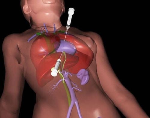
Tim mạch rất quan trọng trong cơ thể người
2. Diagnostic methods for pericardial effusion
2.1 Straight chest radiograph
Normally, the heart-to-thoracic shadow ratio is less than 0.5. When there is fluid in the pericardial cavity, the heart balloon will grow rapidly, bulb-shaped. At the same time, because the blood to the lungs is limited, two pulmonary lung fields are observed to be bright.
This symptom only occurs when the pericardial effusion occurs gradually. Conversely, cardiac shadow may be normal in cases of pericardial effusion causing acute tamponade.
However, it should be noted that fluid less than 250 ml usually does not cause an enlarged heart but can still cause fatal acute cardiac tamponade.
2.2 Electrocardiogram
Electrocardiogram showing signs of pericarditis, pericardial effusion or suggestive of acute cardiac tamponade.
However, as imaging tools have become commonplace, the role of the electrocardiogram in the diagnosis of pericardial effusion has become increasingly limited.
2.3 Echocardiography
This is the most accurate and convenient clinical tool for diagnosing pericardial effusion, in terms of quantity and location of fluid distribution.
The image of the heart dancing (shaking) in the fluid-filled pericardial cavity is the best evidence of pericardial effusion. Besides, the degree of effusion is divided into 4 levels based on the thickness of the fluid layer: small (less than 10 mm), medium (10 - 20 mm), large (20 mm) and very large (over 20 mm). .
Echocardiography is also very necessary in the case of identifying patients with hypotension, cardiovascular collapse due to acute cardiac tamponade.
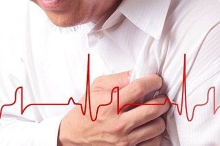
Người bệnh thường biểu hiện triệu chứng khó thở, khó nuốt, ho....
3. Diagnose the cause
Clinical symptoms and signs on echocardiography, electrocardiogram, X-ray as above help to recognize the presence of fluid in the pericardial cavity. The cause of pericardial effusion can only be known by testing the fluid.
Common etiology tests are:
Cancer: Cancer-indicating tests and cytology. Tuberculosis: AFB staining, bacterial culture, .. Germs: MNT fluid culture with at least 3 samples, blood culture must be concurrently with fluid culture. Viruses: PCR testing of some common viruses can help in differential diagnosis from self-reactive pericarditis. Some other tests: Biochemistry: Determine density, protein, LDH, glucose to know whether the fluid is permeable or exudative. Protein, LDH, and blood glucose should be tested at the same time. Blood cell formula: Consists of red blood cells, polymorphonuclear leukocytes, and monocytes. Malignant cells. Cholesterol 3.1 Differential diagnosis
Acute right ventricular myocardial infarction, large pulmonary embolism, aortic dissection are the diagnoses to think about in parallel with the diagnosis of pericardial effusion, cardiac tamponade, and cardiac tamponade. can be excluded.
Since these are all acute cardiovascular diseases, serious complications, life-threatening, the differential diagnosis should be quickly established. Moreover, the treatment direction of each disease is also completely different. Specifically, if it is cardiac tamponade, just need to do pericardial puncture for emergency decompression at the bed, the heart chambers are released, the shock will quickly recover, keeping the patient's life.
To help patients determine the cause, complications and grade of pericardial effusion, Vinmec International General Hospital now has a Cardiovascular Screening Package - Cardiovascular Basic Examination, for all subjects.
The advantages of cardiovascular screening at Vinmec International General Hospital include:
The medical team - doctors are leading experts, highly qualified, dedicated and devoted to the benefit of the patient. patients, bringing high efficiency in medical examination and treatment; Comprehensive and professional medical examination, consultation and treatment services; System of modern equipment, supporting effective diagnosis and treatment; Modern, civilized, luxurious and sterile medical examination and treatment space.
Please dial HOTLINE for more information or register for an appointment HERE. Download MyVinmec app to make appointments faster and to manage your bookings easily.