This is an automatically translated article.
The article was professionally consulted by Specialist Doctor I Tran Cong Trinh - Radiologist - Radiology Department - Vinmec Central Park International General Hospital. The doctor has extensive experience in the field of diagnostic imaging.Appendicitis is an inflammation of the appendix, a closed tube of tissue attached to the large intestine in the lower right abdomen. Inflammation can occur when the appendix becomes infected or blocked by stool, a foreign body, or a tumor.
Your doctor may use an abdominal or pelvic ultrasound, a CT of the abdomen and pelvis, an MRI of the pelvis, or an X-ray to evaluate your condition. The most common treatment for appendicitis is surgical removal of the appendix. If your appendix ruptures and creates an abscess, your doctor may recommend percutaneous drainage of the abscess to remove infected fluid from your body.
1. What is appendicitis?
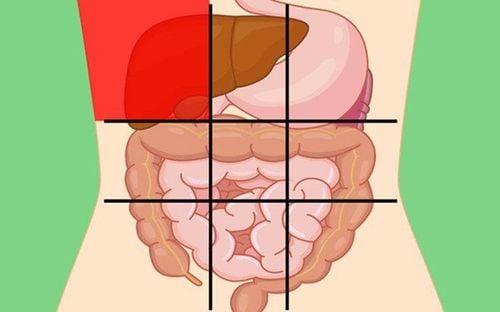
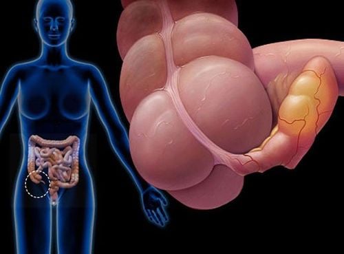
Appendicitis is an inflammation of the appendix, which is a sealed (closed) tube of tissue that attaches to the large intestine in the lower right part of the abdomen. Inflammation occurs when the appendix becomes infected or blocked. Obstruction can be the result of:Stool Foreign body (object or substance introduced from the outside) Tumor An early symptom of acute appendicitis is pain, usually in the center of the abdomen but sometimes in the fossa right pot. The pain may be dull at first, but may become more obvious or severe. Accompanying symptoms may include low-grade (above normal) fever, vomiting, or nausea.
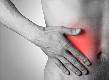
Some individuals, especially children, lose their appetite. As the condition progresses, you will feel severe pain in the lower right part of your abdomen. When the appendix becomes inflamed, symptoms may include:
Severe or worsening pain or pain in the abdomen, rectum, or back Swelling or pain in the abdomen Severe nausea or vomiting High fever Diarrhea or constipation Constipation Inability to defecate.

Sốt cao là một trong những biểu hiện của viêm ruột thừa
2. CT scan, MRI in diagnosis of appendicitis
2.1 CT scan in the diagnosis of appendicitis CT is considered to be a suitable method for the evaluation of appendicitis because it can clearly identify the colon wall as well as the pericolon fat tissue with a sensitivity of 93%. and specificity is 100% in diagnosing or excluding appendicitis. In determining the presence and presence of pericolon complications, CT is also much more sensitive than Baryt's contrast-enhanced colonoscopy.Besides, CT appendicitis is considered to be extremely convenient when used for the purpose of detecting various causes of left lower quadrant pain that can be confused with diverticulitis and may cause diverticulitis. can accurately diagnose up to 78% of cases due to other causes.
The CT images of diverticulitis are correlated with pathologic features.
If appendicitis does not occur in an atypical site, the combination of the presence of diverticula and disproportionately suggestive fatty infiltrates suggests the diagnosis even when occurring in an unusual location. In the presence of: right colon, transverse colon, terminal ileum and jejunum, the diagnosis of diverticulitis by CT scan can be difficult to confirm.
Clinically, cecal diverticulitis can be confused with acute appendicitis. To avoid surgery and prevent possible complications, early and accurate diagnosis is extremely important.
The outstanding advantage of CT scan of appendicitis is its high sensitivity and specificity. In the past decade, with the remarkable advancement of technology, CT scan has created an increasingly high quality appendicitis image. Great.
However, the main disadvantage of CT is radiation problems, especially children and adolescents who are very vulnerable to X-rays. In addition, iodinated contrast material is injected during the procedure. CT scan is very likely to cause allergic reactions and nephrotoxicity. The loss of time to take contrast causes delays in diagnosis and treatment, higher costs. In addition, CT scan results also depend on the skills of performing and interpreting results of technicians and radiologists.
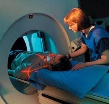
Chụp CT trong chẩn đoán viêm ruột thừa
Currently, MRI is considered feasible and relatively accurate with its sensitivity and specificity for appendicitis up to 85% and 97% respectively. In fact, studies have demonstrated that MRI results in high accuracy and cost savings by patient selection in this trial, which included patients with low to high probability of appendicitis, cost savings can multiply.
Up to now, abdominal magnetic resonance imaging (MRI) is proven to be the most advanced technique in detecting, assessing and monitoring abdominal pathologies including acute appendicitis with the following advantages: outstanding, such as:
This method is non-radioactive, non-invasive, safe for the health of the patient Many types of patients can apply this method The doctor evaluates in detail and clearly the structure of soft tissue inside the abdomen such as liver, kidney, ... based on MRI images The outstanding advantage of MRI for pregnant patients is to avoid radiation exposure to the fetus. pediatric. Performing an MRI is less operator dependent, without the use of intravenous iodinated contrast agents. In addition to diagnosing appendicitis, MRI can also diagnose other conditions such as ovarian torsion or kidney obstruction.
Long execution time is the main disadvantage of MRI. In addition, if the patient has claustrophobia or carries metal devices, MRI is also not possible, the cost of MRI is also relatively higher than other imaging methods.
Besides the methods above, to diagnose appendicitis, the doctor may perform an abdominal or pelvic ultrasound. Ultrasound is a technique that uses sound waves to create images of the inside of the abdomen or pelvis.
In some cases, an abdominal or chest X-ray may be the initial imaging study. Constipation and even pneumonia can cause abdominal pain similar to appendicitis.
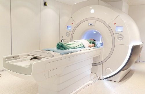
Chụp cộng hưởng từ (MRI) trong chẩn đoán viêm ruột thừa
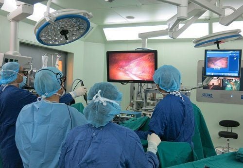
3. Appendiceal Treatment
Appendectomy or surgical removal of the appendix is a common treatment for appendicitis. However, the appendix can rupture for some patients, leading to an abscess or collection of pus. In this situation. To remove fluid from your body in addition to an appendectomy, your doctor may recommend an abscess drainage procedure. You will need to be hospitalized for a few days if the abscess is drained. To ensure that healing goes according to plan, follow-up is usually done on an outpatient basis and you will be attended by a radiologist.Vinmec International General Hospital with a system of modern facilities, medical equipment and a team of experts and doctors with many years of experience in medical examination and treatment, patients can rest assured to visit. examination and treatment at the Hospital.
Please dial HOTLINE for more information or register for an appointment HERE. Download MyVinmec app to make appointments faster and to manage your bookings easily.
Reference source: radiologyinfo.org