This is an automatically translated article.
The article is professionally consulted by Doctor Department of Medical Examination & Internal Medicine - Vinmec Hai Phong International General Hospital.
Appendicitis is a common surgical emergency, accounting for 60-70% of emergency abdominal surgery. The risk of appendicitis per person according to the literature is 7%. Early diagnosis and surgery are decisive factors for good treatment outcomes and minimal complications.
1. General information about appendicitis
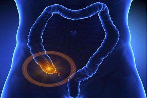
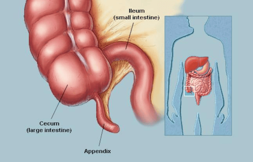
The appendix is a part of the body, a small, thumb-shaped tube located on the right side of the abdomen. One end is sealed off and the other end is connected to the cecum (first segment of the large intestine). When the appendix is blocked, it can be caused by a number of reasons such as: kidney stones, foreign bodies ... it is easy to become inflamed, swollen, and infected, causing the appendix to become inflamed.
Acute appendicitis is the most common surgical emergency in which 70% of cases of acute appendicitis have a typical clinical presentation. The highest incidence is in the age group of 20 - 30 years. The diagnosis of acute appendicitis rests with the clinician. However, for atypical cases, imaging plays a certain important role.
A recent study showed that, based on clinical alone, the diagnosis of acute appendicitis can be wrong up to 30%, combined with ultrasound, this rate can be reduced by up to 15%, and if this method is used With computed tomography, the error rate can be reduced to 7%. The mortality rate of acute appendicitis is about 1% and is directly related to complications from ruptured appendix because it is not properly diagnosed and managed promptly.
Appendicitis is caused by obstruction in the lumen of the appendix due to many causes, including fecal stones 35%, hypertrophy of submucosal lymph cysts 60%, foreign bodies 4% and tumors of the intestine. excess or cecum is 1%. This obstruction increases the pressure in the appendix lumen due to bacterial growth that causes infection and increased secretion of the inner lining of the appendix. As a result, the appendix becomes inflamed, swollen, and filled with pus. If not treated promptly and properly, the appendix can gangrene, or rupture, causing widespread inflammation in the abdomen called peritonitis.
2. The role of ultrasound in the diagnosis of appendicitis
Ultrasound should be chosen as the first imaging modality performed, especially in adolescents, young adults, predisposing subjects of appendicitis, as well as pregnant women. A positive ultrasound result indicates an inflamed appendix. Ultrasonography does not show an inflamed appendix or cannot identify the appendix, with a high suspicion of clinical appendicitis, then a CT scan, or an MRI, should be performed in the pregnant woman. If the patient still has persistent symptoms, especially if the initial ultrasound is performed by someone with little experience in imaging, a repeat ultrasound is a reasonable alternative to a CT scan.
Acute appendicitis is the most commonly thought of emergency diagnosis in patients with acute abdominal pain, and is the most common indication for urgent surgical abdominal intervention. However, it is difficult to make a diagnosis based on history alone, physical examination, and blood and biochemical tests. Many diseases of the gastrointestinal tract, urogenital tract have symptoms quite similar to appendicitis. In pregnant women, appendicitis is the most common cause of abdominal pain requiring surgery. Delayed and unnecessary interventions can have adverse consequences for the fetus.
According to reports, when the diagnosis is based only on clinical and blood and biochemical tests, the average rate of laparotomy without appendicitis is 26% (16 - 47%), while if there are other With imaging methods, this rate is reduced to 6 - 10%. Therefore, the use of imaging methods is very important to confirm the diagnosis when appendicitis is clinically suspected.
Ultrasound examination technique to evaluate the appendix was first mentioned by the author Puylaert J.B.C.M in 1986, since then the ultrasound technique has played an effective role in the diagnosis and treatment of inflammation. acute appendicitis. In this technique, the doctor uses a high-frequency transducer and then gradually and gently presses the transducer over the painful sensitive area suspected of having an inflamed appendix to examine and evaluate the swelling of the inflamed appendix. . Thus, the slow transducer compression technique has three main purposes:
Push the air inside the small intestine and the small bowel loops themselves out of the area to be examined. The optimal approach to the appendix is to examine the appendix in the high-frequency beam focal region of the transducer. Evaluation of the swelling of the inflamed appendix. Ultrasound is valuable because detecting appendicitis has a sensitivity of up to 98.5%, a specificity of 98.2%, and a positive predictive value of 98%. In addition, it is also very valuable to diagnose other diseases causing pain in the right iliac fossa, especially gynecological and urological diseases.
2.1 Normal appendix image Normal appendix arises from cecum, tubular structure, no peristalsis, from outside to inside, including serosa, muscle layer, submucosa, mucosa and lumen appendix, the wall is about 2mm thick and less than 6-7mm in diameter. (Figure 1).
The appendix has a tubular, bowel-like structure (arrow), 2 mm thick wall, 4.5 mm wide (Figure b) seen anterior to the right common iliac artery on sonography. A normal appendix on ultrasound helps to diagnose appendicitis. No further imaging studies were performed.
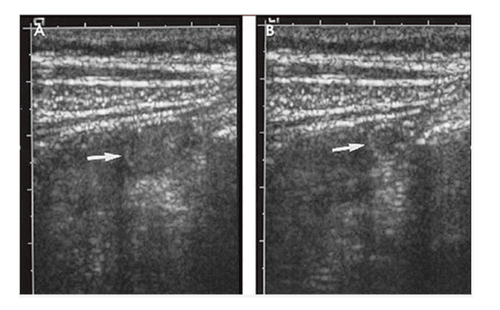
2.2 Image of appendicitis Inflamed appendix on ultrasound is a tubular, fluid-filled, bowel-like structure that is non-compressible, thick-walled, and larger than 6-7 mm in diameter. . Using a transducer, it is possible to distinguish a movable loop of bowel from a fixed inflamed appendix.
Color and power Doppler shows the wall of a tubular structure with hyperperfusion.
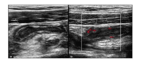
Figure (a) longitudinal section gray-scale ultrasound. Tubular, fluid-filled, bowel-like, non-compressible, thick-walled, and larger than 6-7 mm in diameter, located in the right lower quadrant (arrow).
Figure (b) Power Doppler ultrasound. The structural wall shows increased perfusion (arrow). According to the diagnosis of acute appendicitis on ultrasound, the patient should be operated on, without any further imaging studies.
2.3 Appendicitis at the apex Manual pressure into the lumbar fossa can show appendicitis more clearly, especially when it is posterior to the cecum. The entire appendix should be visualized if the inflammation is localized to the end of the appendix, which is termed “apical” appendicitis. When there is clinical suspicion of appendicitis, the presence of fecal stones in the appendix helps confirm the diagnosis. (Figure 3).
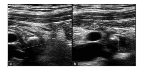
(a) Long-axis view: The appendix was normal at the proximal end, 4.8mm wide, then enlarged, 12.7mm wide, containing fluid at the distal appendix. An echogenic structure, with posterior echogenic shadow consistent with fecal stones, was found within the lumen of the appendix, near the enlarged appendix. Diagnosis of inflammation at the apex of the appendix with fecal stones in the appendix, confirmed postoperatively.
(b) Short axial view: The appendix was normal at the proximal end (4.6mm) and inflamed at the distal (13.9mm), the terminal part contained fecal stones.
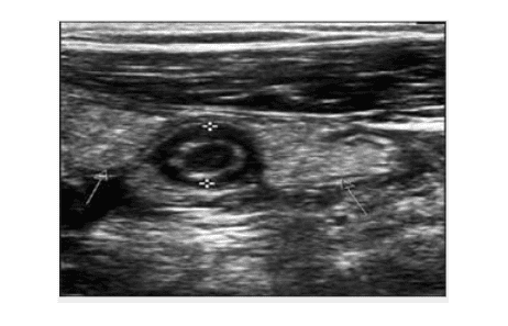
2.4 Image of intra-abdominal fluid Detecting more fluid in the abdomen, between bowel loops and in the iliac fossa, the inflamed mesenteric fat around the appendix is hyperechoic (Figure 4).
In some cases, ultrasound diagnosis of acute appendicitis may be difficult such as:
Patient is obese – ultrasound waves are difficult to penetrate deeply. Paralytic ileus or small bowel obstruction, which is common in acute appendicitis with slightly obscure complications, limits the investigation. Abdominal wall reaction – difficult to perform ultrasound technique (difficulty in pressing the transducer asymptotically with appendicitis). The bladder is too distended, or the patient is more than 6 months pregnant – difficult to perform the compression technique. Abnormal appendix position. The sonographer is inexperienced. Through this article, it is clear that the role of ultrasound in the diagnosis of appendicitis. Ultrasound is a non-traumatic imaging method, capable of being performed quickly in all medical facilities, potentially contributing to increasing the rate of correct diagnosis of acute appendicitis. Ultrasonography is also useful in the differential diagnosis of other gastrointestinal diseases such as ileocecal inflammation, mesenteric lymphadenitis, cecal diverticulitis, and gynecological or urological conditions.
Vinmec International General Hospital is one of the hospitals that not only ensures professional quality with a team of doctors, modern equipment and technology, but also stands out for its examination, consulting and service services. comprehensive and professional medical treatment; civilized, polite, safe and sterile medical examination and treatment space.
Please dial HOTLINE for more information or register for an appointment HERE. Download MyVinmec app to make appointments faster and to manage your bookings easily.