This is an automatically translated article.
Posted by Doctor Mai Vien Phuong - Department of Examination & Internal Medicine - Vinmec Central Park International Hospital
Cytoscopy provides a high-quality, time-saving, low-cost diagnostic procedure, capable of detailed detection, characterization of gastrointestinal tumors, determining the extent of tumors. transform cancer cells by visualization of cell size changes, polarization disturbances, and nuclear distortions.
1. Cell endoscopy with the help of artificial intelligence
In the past few years, new approaches have emerged in endoscopic imaging technology for the high-quality assessment of lesions found in the gastrointestinal (GI) tract.
Cytoscopy is a new tool in endoscopy, which helps to more accurately evaluate the superficial mucosal surface. Endoscopic cytology provides a high-quality, time-saving, low-cost diagnostic procedure, capable of detailed detection, characterization of gastrointestinal tumors, and determination of the degree of variation. modifying cancer cells by visualizing cell size changes, polarization disturbances, and nuclear distortions.
In particular, the cyto endoscopy system can assess the depth of cancer invasion and predict the outcome of treatment. Along with that, one of the significant benefits of cytoscopy is that it is powered by artificial intelligence (AI), which ensures enhanced delineation between benign and cancerous digestive lesions.
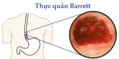
>>> Diagnosis of Barrett's esophagus and esophageal cancer by artificial intelligence

2. Real-time cyto endoscopy
In the last few years, there has been increasing interest in the application of computer-aided diagnostics (CAD) systems as a new tool in improving the quality of cytology.
The diagnostic algorithm of the EC-CAD cyto endoscopy system includes 3 main steps: nuclear segmentation, mucosal feature extraction and prediction pathology classification output.
The main benefit of using the EC-CAD cyto endoscopy system is its high diagnostic capabilities “in real time” during endoscopy. More recent studies have shown that the correlation between EC-CAD cytology and gastrointestinal pathology findings is lower. This could allow this diagnostic endoscopic approach to serve as a kind of in situ “optical biopsy”.

3. Several generations of cytoscope
The first generation cytoscope was introduced into clinical practice in 2003 (XCE120U; Olympus Medical Systems Corp., Tokyo, Japan). Subsequently, an improved version of cytoscope with a dual integrated type lens was introduced in 2005 (gastrointestinal F-Y0001; Olympus Medical Systems Corp., Tokyo, Japan). 4 years later, in 2009, the 3rd generation of cytoscope appeared with a single integral type lens and smaller outer diameter (gastrointestinal F-Y0002; Olympus Medical Systems Corp., Tokyo, Japan). The latest version of cytoscopy emerged in 2015 with the ability to provide high quality colonoscopy (gastrointestinal F-H290CEI; Olympus Medical Systems Corp., Tokyo, Japan).
>>> Application of artificial intelligence in the treatment of pancreatic cancer
This latest model of cytoskeletal endoscopy includes Magnified-NBI observation and cyto endoscopy with 520x magnification. With the exception of integrated cytoscope, in clinical practice there exists a new model of probe-based cyto endoscopy with higher magnification (1390x), which provides simultaneous acquisition of biopsies from area of interest.
In 2019, Olympus introduced new real-time cytoreductive imaging, based on artificial intelligence software. This new development is called "Endobrain" and could be applied to conventional colonoscopy as a real-time diagnostic aid, allowing direct treatment decisions.
4. Steps to prepare stomach and colon before cytososcopy
Cytoscopy requires good mucosal preparation to provide detailed images of colonic lesions. After rinsing the mucosa with water, the second preparation step is to use simethicone and N-acetylcysteine (a dye solution for staining the mucosa). Based on the literature, 3 dyes with different concentrations exist, methylene blue (MB), toluidine blue and crystal violet (CV). In 2006, Kodashima et al published a procedure using 0.25% toluidine blue in the stomach and colon, with an exposure time of 60 seconds. According to Ichimasa et al., a mixture of 1% MB and 0.05% CV for colonoscopy is superior to other staining combinations.

5. The role of AI-assisted cytoscope in colorectal cancer
With the aid of artificial intelligence in cytoscopy, treatment options for colonic lesions become more accessible and accurate. Artificial intelligence technology provides "real-time" histology, so it is possible to determine whether a sizable colon lesion (>2 cm) should be treated with surgery or laparoscopic resection . Artificial intelligence endoscopy significantly shortens the process of making a histological diagnosis and avoids unnecessary tissue biopsies.
Lui et al supported a study to evaluate the application of an AI-assisted image classifier to determine the feasibility of laparoscopic resection for these lesions. large colonic lesions based on endoscopic images without magnification. They collected an artificial intelligence image classifier using 8000 endoscopic images of colonic lesions. Meanwhile, the validation set included 567 endoscopic images from 76 patients. The histological findings of resected specimens were used as the gold standard for validation in the study.
>>> Artificial intelligence and gastric cancer
Therapeutic endoscopic resection is performed only in patients with well-differentiated adenocarcinoma, with submucosal invasion 1mm as well as no any lymphatic invasion. The results obtained by the artificial intelligence image classifier are compared with those captured by (experienced) endoscopists. In patients with previously mentioned lesions, who were indicated for laparoscopic surgery, artificial intelligence had excellent accuracy (85.5%). This study highlights the clinical significance of artificial intelligence in predicting laparoscopic resection of large colonic lesions (>2 cm).
Although the gold standard for histological observation of gastrointestinal lesions is usually based on light microscopy analysis of hematoxylin and eosin-stained slice specimens. However, real-time endoscopic cytological evaluation can also aid in histopathology, allowing detection of lesions at the cellular level and assessment of cell loss. Both methods showed a significant correlation. Artificial intelligence-assisted cytoreduction is gradually changing the approach of medicine to the treatment of gastrointestinal lesions. In fact, not all lesions detected by colonoscopy need to be removed or sent for histopathological evaluation.
6. Conclusion
Before AI-assisted cytoscopy became a common practice, significant barriers such as patient acceptance or performance by poorly qualified endoscopists and Regulatory issues need to be handled with care. The development of CAD algorithms and artificial intelligence can promote, shape and improve decision making in the management of colorectal lesions. Overall, cytology has shown excellent diagnostic accuracy, helping to aid in the rapid diagnosis of lesions in the lower gastrointestinal tract.
Currently, Vinmec International General Hospital is a prestigious address trusted by many patients in performing diagnostic techniques for digestive diseases, diseases that cause chronic diarrhea or Crohn's disease... In particular, the screening techniques for gastric cancer and gastric polyps at Vinmec were performed using the Olympus CV 190 endoscope, the NBI function (Narrow Banding Imaging - endoscopy with narrow light frequency band) for image results. analyze mucosal pathology more clearly... Thanks to that, the doctor can make an accurate diagnosis and give a timely treatment plan. Not only has a modern equipment system, Vinmec is also a place to gather a team of experienced doctors and nurses, especially, with a space designed according to 5-star hotel standards, Vinmec is guaranteed to bring bring the patient comfort, friendliness, peace of mind.
Please dial HOTLINE for more information or register for an appointment HERE. Download MyVinmec app to make appointments faster and to manage your bookings easily.
References
1. Neumann H, Fuchs FS, Vieth M, Atreya R, Siebler J, Kiesslich R, Neurath MF. Review article: in vivo imaging by endocytoscopy. Aliment Pharmacol Ther . 2011; 33 :1183-1193. [PubMed] [DOI]
2. Sasajima K , Kudo SE, Inoue H, Takeuchi T, Kashida H, Hidaka E, Kawachi H, Sakashita M, Tanaka J, Shiokawa A. Real-time in vivo virtual histology of colorectal lesions when using the endocytoscopy system. Gastrointest Endosc. 2006; 63 1010-1017. [PubMed] [DOI]
3. Takamaru H , Wu SYS, Saito Y. Endocytoscopy: technology and clinical application in the lower gastrointestinal tract. Transl Gastroenterol Hepatol . 2020; 5:40. [PubMed] [DOI]
4. Inoue H , Kudo SE, Shiokawa A. Technology insight: Laser-scanning confocal microscopy and endocytoscopy for cellular observation of the gastrointestinal tract. Nat Clin Pract Gastroenterol Hepatol . 2005; 2:31-37. [PubMed] [DOI]
5. Kudo SE , Wakamura K, Ikehara N, Mori Y, Inoue H, Hamatani S. Diagnosis of colorectal lesions with a novel endocytoscopic classification - a pilot study. Endoscopy . 2011; 43 :869-875. [PubMed] [DOI]


