This is an automatically translated article.
The article was professionally consulted by Specialist Doctor I Tran Cong Trinh - Radiologist - Radiology Department - Vinmec Central Park International General Hospital. The doctor has many years of experience in the field of diagnostic imaging.Abnormal changes of heart shadow on X-ray are a warning sign of abnormal heart disease such as: Large, small heart shadow; Large left atrium, large right atrium... Abnormal images of the heart shadow on radiographs can help doctors orientate a number of cardiovascular pathologies, thereby giving subclinical indications and accurate diagnosis. and appropriate treatment.
1. Big heart ball
To determine the cardiac shadow on chest X-ray, it is necessary to determine the cardiothoracic ratio, the cardiothoracic index is the ratio between the horizontal size of the heart shadow and the width of the chest cavity. A normal cardiac shadow on straight chest radiographs has a cardiothoracic ratio of 0.5-0.55. The heart shadow is enlarged when the heart-to-chest ratio is greater than 0.55.Causes of enlarged heart on radiographs include:
Heart failure: Heart failure can be caused by many causes, when there is heart failure, the heart muscle cells must increase their work capacity, leading to The heart muscle is enlarged, blood is pooled in the heart chambers and forms a large shadow on the chest X-ray. Heart valve disease: An abnormality of the heart valve system such as: stenosis of the mitral valve, tricuspid valve or aortic valve... Also causes blood to pool in the heart chambers, causing a large heart balloon on the film. . Pericardial effusion: Pericardial effusion is the cause of clinical shortness of breath and chest pain. Pericardial effusion is an abnormal increase in fluid between the pericardium, leading to cardiac tamponade. The phenomenon of increased fluid in the abnormal pericardium also changes the image of the heart shadow on the X-ray film. Congenital heart disease such as: From fallot (heart-shaped like a boot), Ebstein's disease (heart shaped like a box)...
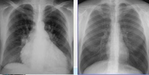
Hình ảnh bóng tim to trên X quang
2. Small heart ball
When the thorax elongates, the heart is in a suspended state. At that time, the axis of the heart was almost parallel to the vertical axis of the body, it was called a teardrop-shaped heart, and the diameter of the heart decreased markedly. Small heart balloon condition can be seen in diseases such as:Emphysema caused by dysfunction of alveolar sacs and bronchioles due to overstretching or destruction by chronic inflammatory process count. This situation lasts for a long time, causing the respiratory system to lose elasticity, after inhaling, the patient has difficulty breathing in the exhalation, so the air is trapped in the lungs, forming air sacs containing oxygen-poor air. A typical chest x-ray shows an emphysema and a teardrop-shaped heart shadow. Chronic lung disease, bronchial asthma: This condition causes gas to accumulate in the lungs, so the chest X-ray film is wide in the shape of a barrel, the heart shadow is small. Anemia, physical exhaustion: On the X-ray film, there is a small chest image and a small heart shadow.

Hen phế quản là nguyên nhân gây ra hiện tượng bóng tim nhỏ trên phim chụp
3. Large left atrium
Image of heart shadow on X-ray film straight and inclined, can see the condition of left atrial enlargement. Large left atrium can be caused by:Patient has stenosis and mitral regurgitation: The mitral stenosis causes the right atrium to increase contraction to push blood down to the left ventricle, the amount of blood in the left atrium is congested. cause left atrial enlargement. Mitral regurgitation causes blood from the left ventricle to pump back into the left atrium during contraction, increasing the amount of blood in the left atrium leading to a large left atrium image on radiographs. Left atrial enlargement depends on the degree that manifests differently on the film. Heart failure: Heart failure is often accompanied by an enlarged heart.
4. Large right atrium
Right atrial enlargement is often caused by: congenital heart disease, mitral and tricuspid valve disease, chronic valvular heart disease...Right atrium enlargement on X-ray is determined by the size of the Right atrium, if the size of the right atrium is greater than 5.5 cm, then the right atrium is considered large.

Trẻ mắc bệnh tim bẩm sinh thường có hiện tượng lớn nhĩ phải
5. The right heart is big
Large right ventricular image can show enlarged lung hilum; On slanted film, a loss of light in front of the heart can be seen.Causes of right ventricular enlargement include:
Pulmonary stenosis: This condition causes blood to pool in the right ventricle leading to right ventricular dilatation, causing right ventricular enlargement. Mitral stenosis: In case of mitral stenosis, the accompanying sign on the radiograph is an enlarged left atrium. Pulmonary diseases: Some lung diseases increase blood vessel pressure in the lungs, leading to increased pressure on the right ventricle. This leads to an enlarged right ventricle. Cardiopulmonary X-ray film technique is a simple technique that helps to survey abnormalities of organs in the chest such as muscles, bones, lungs, and heart. On the film can see abnormal images of the heart shadow, from which images can help guide diagnosis, diagnosis and follow-up treatment of the disease.
Please dial HOTLINE for more information or register for an appointment HERE. Download MyVinmec app to make appointments faster and to manage your bookings easily.
SEE MORE
Diseases that can be diagnosed early by chest X-ray Do X-rays affect health? Radiation level when taking X-ray


