This is an automatically translated article.
The article is professionally consulted by Master, Doctor Tong Diu Huong - Radiologist - Department of Diagnostic Imaging - Vinmec Nha Trang International General Hospital.X-ray of the file is a technique to find the cause of pain in the pharynx, which is suspected to be caused by an overgrown file, and is indicated in cases where the patient has symptoms such as difficulty swallowing, painful swallowing. ..
1. What is brooch length?
In the human body, the brooch is the one-headed bone that attaches to the temporal bone, the normal brooch will be less than 2.5cm in length, if it's over 3cm, it's considered long.
Long brooch is the phenomenon where the brooch is stretched too long and causes certain discomfort to the patient in the throat and ear area. The disease was first described in 1937 by Dr. Watt Eagle, the common sign of long styloid in patients is difficulty swallowing, painful swallowing, palpation of the tonsil fossa shows the tip of the file.
2. Indications and contraindications for styrofoam X-ray
X-ray of the file is a method that is used quite commonly:
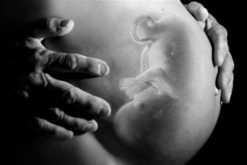
Indication for X-ray of the file: The patient has signs of difficulty swallowing, painful swallowing, palpating the tonsil fossa can see the tip of the file. brooch: There are no absolute contraindications, relative contraindications for pregnant women

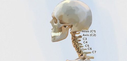
Vị trí mỏm trâm trên hình ảnh giải phẫu
3. X-ray procedure of the file
Step 1: The technician prepares supplies, the patient removes jewelry such as necklaces, earrings, hairpins (if any)
Step 2: The patient lies supine on the X-ray table so that the plane is flat. the front is perpendicular to the film and the arms are stretched out comfortably. The median line and the ear are perpendicular to the film, the patient opens his mouth. In the case of lying on the stomach, the two hands must be folded and propped up on the table.
Step 3: Place the sand-test
Step 4: Correct the X-ray shadow
Step 5: Set the imaging constant
Step 6: Carry out the scan
Step 7: Guide the patient out of the room. take the film to be washed or printed for the CR/DR system.
X-ray technique is quite simple and has no complications, however, if the patient encounters errors such as not being able to keep still during the film, the image will not be clearly revealed. and have to take a picture.

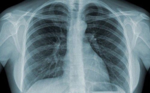
Kết quả X quang mỏm trâm dài
X-ray techniques as well as many other medical examination and treatment techniques at Vinmec International General Hospital are conducted by a team of highly qualified and experienced medical doctors; the system of advanced and modern machinery should significantly reduce radiation dose with the best image and always be updated according to medical progress in the world; Especially, professional service quality will help customers have the most comfortable and secure experience when visiting Vinmec. Customers who need advice and support, please contact HERE.
SEE MORE
Relationship between X-rays and Pregnancy In what case is X-ray of the uterus and fallopian tubes (HSG scan) done? Mammomat Inspiration X-ray machine - an effective "assistant" in the diagnosis and treatment of breast tumors