This is an automatically translated article.
The article is expertly consulted by Master, Doctor Trinh Thi Phuong Nga - Radiologist - Department of Diagnostic Imaging and Nuclear Medicine - Vinmec Times City International General Hospital.The carotid and vertebral arteries are important vascular systems responsible for supplying blood and oxygen to the brain. Diseases related to the carotid artery such as narrowing or atherosclerosis can cause stroke and death. Carotid ultrasound is an effective method often used by doctors to examine the condition of the carotid system.
1. What is carotid and vertebral artery disease?
The carotid artery is a blood vessel that rises from the thoracic aorta to nourish the brain. The carotid artery consists of segments of the common carotid artery and then splits into two branches of the internal carotid artery and the external carotid artery in the neck above the thyroid cartilage, across the 4th cervical vertebra.Atherosclerotic plaques form in the wall. The carotid artery reduces the amount of blood to the brain, creating a very dangerous carotid artery disease. If not detected and treated in time, it will cause a stroke and lead to death.
The older you get, the higher your risk of carotid artery disease. In people under 60 years old, the rate is only 1%, but in the elderly over 80 years old, the rate of carotid artery disease is up to 10%.
2. What is carotid and vertebral artery ultrasound?
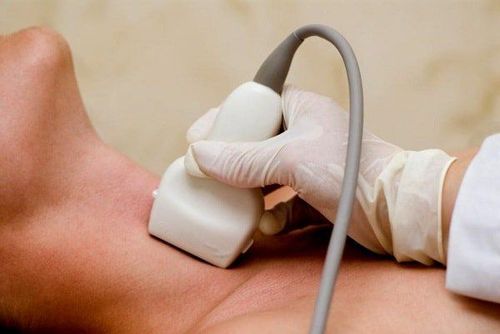
Ultrasound of the carotid and vertebral arteries is an easy, fast, convenient and accurate imaging technique to image the blood vessels in the neck.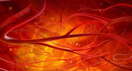
Siêu âm mạch cảnh giúp chẩn đoán hình ảnh các mạch máu ở cổ
Carotid and vertebral artery ultrasound is an easy, quick and low-cost method to perform and is the first choice of specialists when there is a suspicion of carotid artery disease.
3. When does the doctor order an ultrasound of the carotid and vertebral arteries?
The doctor will appoint the patient to perform an ultrasound of the carotid artery and vertebral artery in the following cases:● The patient has signs of a mild, transient stroke or a history of stroke, signs of an ear attack. Mild changes may appear for no more than 24 hours and disappear soon after. It could be all of the symptoms of a stroke or some of it.
● The doctor hears abnormal carotid sounds during the examination.
There are suspicions of carotid artery wall weakness, bulging or dissection, atherosclerosis, or occlusion of the vertebral artery.
● Indicated to perform before and after surgery involving the aorta, carotid artery, coronary artery, peripheral artery...
● This method can be performed for large patients age, people have a high risk of stroke as a way of disease prevention, especially for adults over 70 years old with diseases such as: Diabetes, high blood pressure, atherosclerosis, dyslipidemia ...

Siêu âm mạch cảnh được chỉ định cho cả bệnh nhân lớn tuổi có bệnh nền
● Patients with transient ischemia.
Transient blindness may be caused by carotid stenosis.
4. Procedure for performing carotid ultrasound
Usually, ultrasound in general and ultrasound of the carotid artery and vertebral artery in particular are painless and take a short time of about 20-30 minutes. Steps to perform carotid and vertebral artery ultrasound are as follows:4.1 Prepare the patient The patient can be changed to perform the ultrasound easily, remove the neck jewelry according to the procedure. directed by Doctor.
The performing doctor will explain to the patient the ultrasound process so that the patient can coordinate smoothly, avoiding wasting time for both.
4.2 Perform carotid and vertebral artery ultrasound The patient lies on the ultrasound bed with the doctor's position, there will be a pillow to help the patient's neck slope down so that the long carotid area can be exposed. possible.
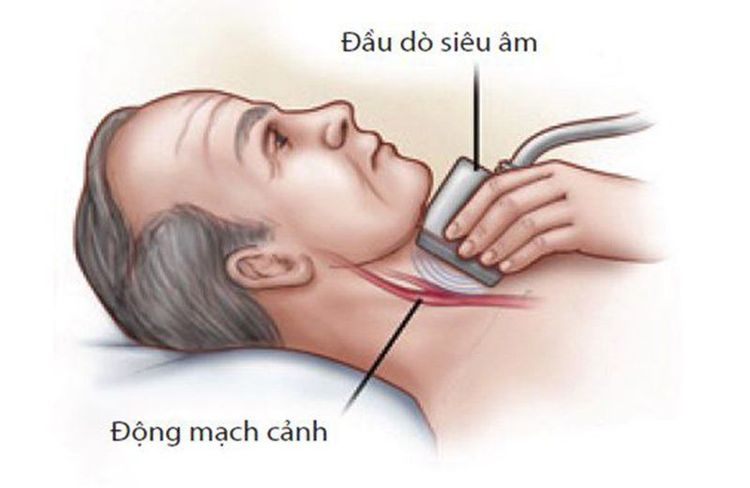
Cổ bệnh nhân dốc xuống sẽ giúp lộ vùng mạch cảnh được dài nhất
The doctor will ask the patient to turn or tilt the direction so that it is convenient for the doctor to do.
Clean the patient's neck and complete the ultrasound.
4.3 Read the results to the patient After the ultrasound is done, the doctor will read the ultrasound results to the patient, inform the patient about the condition of the carotid artery, the vertebral artery and the treatment direction if there is a pathology. some.
Currently, one of the imaging methods to assess carotid and vertebral artery disease with high accuracy and reliability is Doppler ultrasound of the carotid and vertebral arteries. This method can be applied to all subjects, does not affect health and is being applied at Vinmec International General Hospital. This is also one of the medical facilities that put prestige and quality on the top, converging many good specialists.
Doctor Trinh Thi Phuong Nga is a radiologist with nearly 20 years of experience in diagnostic imaging. Currently, the doctor is working at - Department of Diagnostic Imaging - Vinmec Times City International General Hospital.
Any questions that need to be answered by a specialist doctor as well as customers wishing to be examined and treated at Vinmec International General Hospital, you can contact Vinmec Health System nationwide or register online HERE.
LEARN MORE
Why can carotid artery stenosis cause a cerebrovascular accident? How dangerous is carotid stenting? Learn about pseudoaneurysms


